

50th ARGENTINE CONGRESS OF COLOPROCTOLOGY
OFFICIAL REVIEW
ANASTOMOTIC LEAK
AFTER COLORECTAL SURGERY: DIAGNOSIS AND TREATMENT
Mariano Laporte, MD
Coordinator, Colorectal Surgery Unit, General Surgery Department, Sanatorio Güemes
Coordinator, Endoscopy Center, Institute of Digestive Diseases, Hospital Alemán de Buenos Aires
General Surgery Staff, Clínica Bazterrica
Contributors: Agustina Casas, MD; Belén Dening, MD; Ana Leone, MD; Macarena Lucas, MD
Colorectal Surgery Division, General Surgery Department, Sanatorio Güemes
ACKNOWLEDGEMENTS
First and foremost, I sincerely thank the members of the Executive Committee of the Argentine Society of Coloproctology for appointing me to present the Official Review at the Annual Congress of Coloproctology. I am deeply honored to have been selected by colleagues I hold in the highest regard, both for their medical expertise and their personal qualities.
From the beginning of my career, I developed a particular interest in anastomotic complications in colorectal surgery. This subject has motivated me to publish several articles and book chapters and forms the central focus of this Official Review
It is inevitable to look back and reflect on the path I have traveled in a surgical specialty that I am truly passionate about. First, I would like to thank the physicians of the Department of General Surgery at Hospital Posadas, where I completed my residency exactly 20 years ago. In particular, I would like to thank Dr. Carlos Zorraquín, who instilled in me a passion for this specialty. I also have fond memories of my residency colleagues, with whom, fortunately, I have remained in close contact.
I subsequently had the opportunity to complete my training in Coloproctology at Hospital Alemán de Buenos Aires, sharing that period with Dr. Gerardo Zanoni, whom I hold in the highest regard. In the years that followed, serving as Chief Resident in General Surgery and later as an associate staff surgeon in the Department of General Surgery at Hospital Alemán contributed decisively to my professional growth. I am deeply grateful to all the physicians in the Department of General Surgery and, in particular, to the surgical residents of those years, with whom I have developed unique bonds that remain strong to this day.
During my time at Hospital Piñero, I fondly remember Dr. Guillermo Obregón and the residents of that period. Some of them subsequently chose colorectal surgery as their specialty, allowing us to maintain a close relationship over the years. Two years ago, I was invited to return to Sanatorio Güemes and join the Department of General Surgery, led by Dr. Gustavo Castagneto. There, we have had the opportunity to establish and consolidate a Colorectal Surgery Unit together with two highly talented specialists with tremendous potential, Dr. Agustina Casas and Dr. Ana Leone. Along with the current fellows, Dr. Belén Dening and Dr. Macarena Lucas, they have actively contributed to the preparation of this Official Review. I am deeply grateful to all of them.
I would also like to express my sincere appreciation to the residents in the General Surgery Residency Program at Sanatorio Güemes. They play an indispensable role: they provide support, solve problems, learn, teach, and sustain a substantial part of our daily clinical activity. Their commitment, energy, and dedication not only contribute to patient care but also to the academic, educational, and scientific growth of our department.
I would also like to express my sincere gratitude to my colleagues and friends at the Argentine Society of Coloproctology. Over time, I believe we have come to understand that friendship stands above all else. I have chosen not to mention each of them individually, out of concern that I might inadvertently leave someone out.
I would also like to sincerely thank the editors and members of the editorial team of the Revista Argentina de Coloproctología, who devote many hours of their time to promoting and strengthening our journal. I am particularly grateful for their corrections, suggestions, and revisions to this Official Review.
I would like to thank Claudio Rodríguez, from the library at Hospital Alemán, for his continued assistance in accessing the medical literature and, especially, for his invaluable contribution to the literature search for this review.
I am also deeply grateful to those who have always supported me professionally, particularly Dr. Juan Pablo Muñoz and my colleagues in the Department of Surgery at Clínica Bazterrica. I would like to make special mention of my colleagues and the nonmedical staff of the Endoscopy Unit at Hospital Alemán for their daily support, collaboration, and commitment.
It would be impossible not to acknowledge the fundamental role that three colleagues whom I deeply admire have played in my professional training: Dr. Nicolás Rotholtz, Dr. Maximiliano Bun, and Dr. Alejandro Canelas. To them, my most sincere gratitude. It has been a privilege to share so many years of our professional and personal lives. Without a doubt, they have had a decisive influence on my training and on the way I understand surgery, as a practice that demands technical excellence, unwavering commitment, and a profound sense of humanity. I am also grateful for their guidance and for their comments and revisions regarding this Official Review.
I am deeply grateful to my family, especially my mother, Marta, and my brother, Luis, who remind me every day of the importance of moving forward, even in the face of life's challenges and whatever obstacles may arise. To my father, wherever he may be, I offer my heartfelt gratitude for always encouraging me to grow and strive for excellence, and for teaching me by his example that perseverance, dedication, and hard work are the path to achieving one's goals.
Finally, to my wife, Gabriela, who has undoubtedly been a fundamental pillar in my ability to grow and develop in this wonderful specialty. Thank you for raising our three children, Agustina, Juani, and Martín, with such love, generosity, and strength, especially during the many periods when the demands of my profession kept me away from home.
I. Introduction
Anastomotic leakage (AL) remains one of the most feared and challenging complications of colorectal surgery. Despite advances in surgical techniques, new suturing and stapling devices, enhanced recovery protocols, and a better understanding of patient- and procedure-related risk factors, the incidence of AL has remained relatively unchanged over the past several decades.
The pathophysiology of AL is complex and multifactorial. Anastomotic integrity depends on the interplay of patient-related factors, the underlying disease, surgical technique, tissue perfusion, inflammatory responses, and the healing process. More recently, the potential role of host–microbiome interactions has also emerged as a modulator of the local inflammatory response and anastomotic healing. This complex pathophysiology may explain why, despite substantial advances in perioperative care and surgical technique, complete prevention of AL remains elusive.
A comprehensive understanding of the mechanisms underlying anastomotic healing, predisposing factors, available diagnostic modalities, and therapeutic strategies is essential to optimize clinical decision-making and improve patient outcomes.
The clinical significance of AL extends well beyond the initial surgical event. Clinically, AL is a major contributor to postoperative sepsis, reoperation, permanent stoma formation, and mortality. It may also result in significant long-term functional impairment, adversely affecting quality of life and potentially compromising oncologic outcomes.
The aim of this review is to provide a comprehensive contemporary overview of AL in colorectal surgery. The review is organized into sequential chapters addressing the biological mechanisms of intestinal anastomotic healing, the emerging role of the gut microbiome, preoperative and intraoperative risk factors, diagnostic strategies, therapeutic options, and the clinical consequences of AL.
The general goal is to provide colorectal surgeons with a practical, up-to-date, and evidence-based resource.
The structure follows a deliberate logical sequence designed to facilitate a progressive understanding of AL. It begins by examining the biological principles underlying anastomotic integrity and the pathophysiologic mechanisms leading to anastomotic failure, followed by a discussion of prevention, diagnosis, and management, and the impact of AL on patient outcomes.
At the end of each chapter, a conceptual summary integrates the main topics discussed, highlights the key aspects, and facilitates their understanding and translation into clinical practice.
Historically, research on AL has focused primarily on elucidating its pathophysiologic mechanisms, identifying risk factors, and optimizing preventive and therapeutic strategies. More recently, increasing attention has been directed toward another, less explored consequence of this complication: its impact on the surgical team. AL may affect not only patients but also have substantial emotional, professional, and occupational consequences for the surgeons involved in their care.
To further explore this issue, we conducted an anonymous survey of surgeons from different surgical specialties, specifically examining the impact of AL on the surgical team. This analysis provides insight not only into current approaches to the management of AL but also into a frequently overlooked dimension of this complication: its effect on surgeons. Chapter IX of this review presents and discusses the findings of this survey.
II. Physiology of Intestinal Anastomotic Healing
A thorough understanding of the normal process of intestinal anastomotic healing is essential to elucidate the mechanisms underlying AL. Anastomotic healing is a tightly regulated sequence of events that begins with hemostasis and progresses through inflammation and tissue proliferation before culminating in collagen remodeling and restoration of tissue strength.1 This process is traditionally divided into 3 overlapping phases: inflammatory, proliferative, and remodeling.
1) Inflammatory Phase: The inflammatory phase of wound healing, also referred to as the exudative phase, begins with hemostasis, characterized by platelet activation and aggregation, and the formation of fibrin clots. These clots serve not only to arrest bleeding but also to provide a temporary scaffold for the deposition of early extracellular matrix proteins.
In addition to forming fibrin plugs, platelets that aggregate at the site of mucosal injury release multiple cytokines and growth factors, including platelet-derived growth factor (PDGF), vascular endothelial growth factor (VEGF), and transforming growth factor-beta (TGF-β). This cytokine-rich environment at the site of injury increases the permeability of the surrounding microvasculature, thereby promoting the recruitment and migration of inflammatory cells into the healing tissue.2
After platelet aggregation, neutrophils and subsequently macrophages are the first inflammatory cells to migrate to the site of injury. These cell populations remain predominant within the healing anastomosis throughout the inflammatory phase and perform multiple functions, including phagocytosis of potential pathogens and the release of additional growth factors that promote tissue repair and healing.
2) Proliferative Phase: Following the inflammatory phase, the healing intestinal tissue enters a proliferative phase in which fibroblasts become the predominant cell type. The primary function of fibroblasts is to deposit collagen and replace the initial, transient fibrin clot with granulation tissue. Fibroblasts are initially recruited to the transected intestinal edge by chemotactic factors such as platelet-derived growth factor (PDGF), transforming growth factor-beta (TGF-β), and fibroblast growth factor (FGF), which are released by macrophages. In contrast to the collagen composition of mature intestine, whose extracellular matrix contains approximately 20% type III collagen, early granulation tissue synthesized by fibroblasts contains a significantly higher proportion of this collagen type (30%–40%). This tissue therefore requires remodeling during the subsequent phase of healing.3
Fibroblasts begin to appear toward the end of the inflammatory phase and become the predominant cell type within the wound bed by approximately postoperative day 4. The proliferative phase typically extends for approximately 14 days. As fibroblasts accumulate, the previously predominant neutrophils and macrophages decrease in number and are progressively replaced by lymphocytes. In addition to collagen deposition, this phase is characterized by angiogenesis and re-epithelialization of the intestinal mucosal surface, both of which are stimulated by specific growth factors. Toward the end of this phase, smooth muscle cells and myofibroblasts emerge as the anastomosis gains strength and undergoes contraction, contributing to a reduction in the size of the final scar.1
3) Remodeling Phase: The final and longest phase of anastomotic healing begins approximately 14 days after surgery and continues for several weeks as the wound undergoes reorganization into permanent scar tissue.1
The inflammatory cells that predominate during the early stages of healing further decrease in number, while fibroblasts, as in the proliferative phase, remain the predominant cell type. During this phase, the ratio of the 2 major collagen types is restored toward that of mature tissue, as matrix metalloproteinases (MMPs) degrade type III collagen within the granulation tissue, promoting its replacement with type I collagen. Once tissue remodeling and maturation are complete, type I collagen accounts for approximately 68% of the total collagen content of the intestinal submucosa.4
This remodeling process is largely mediated by MMPs and their tissue inhibitors, which facilitate degradation of the immature extracellular matrix and formation of a more stable scar. However, early MMP overexpression may result in excessive collagen degradation and contribute to anastomotic leakage. Factors such as preoperative ischemia and microbiome activation, particularly through matrix metalloproteinase-9 (MMP-9), may increase this risk. Conversely, MMP inhibition has been shown to improve anastomotic strength in experimental models.5
The resulting stronger collagen matrix is reorganized into compact bundles, while myofibroblast contraction contributes to strengthening the healing wound. Similarly, the vascular networks formed during the proliferative phase undergo further organization into mature vascular networks, and over the course of several weeks to months, the anastomotic tissue progressively stabilizes. Despite the time required to regain strength, however, healed wounds and anastomoses recover only a portion of the tensile strength of uninjured tissue.1,2
The acquisition of tensile strength is not uniform throughout the gastrointestinal tract. Ileal and other small-bowel anastomoses not only regain strength more rapidly but also reach values approaching those of normal tissue at approximately 4 weeks postoperatively. In contrast, colonic anastomoses reach only approximately 75% of normal tissue strength at 4 months. These differences in the healing response between the small intestine and colon may be attributable, at least in part, to greater collagenase activity in the colon, which is associated with a transient decrease in anastomotic strength during the first 3 postoperative days. This phenomenon is more pronounced in the colon than in the small intestine and may help explain the differences in strength recovery between these gastrointestinal segments. Clinically, this greater collagenase activity may also contribute to the higher incidence of anastomotic leakage reported after colonic compared with small-bowel anastomoses.3,6
Among the growth factors involved in wound healing, PDGF, TGF-β, and VEGF are particularly important mediators. The roles of PDGF and TGF-β appear relevant, although experimental evidence regarding their direct effects on anastomotic strength remains conflicting. In contrast, VEGF appears to play a central role by promoting neovascularization, improving oxygen and nutrient delivery to the wound bed, and, in animal models, increasing anastomotic strength.7
Overall, intestinal anastomotic healing is a dynamic and sequential process in which initial hemostasis, the inflammatory response, fibroblast proliferation, angiogenesis, re-epithelialization, and collagen remodeling act in concert to restore anastomotic continuity and strength. Specific features of each phase help explain how factors such as ischemia, local contamination, microbiome alterations, an imbalance between MMPs and tissue inhibitors of metalloproteinases (TIMPs), or inadequate angiogenesis may compromise anastomotic integrity and promote the development of AL. Understanding the sequence and mechanisms underlying normal healing is therefore essential for interpreting the pathophysiologic mechanisms involved in AL.
CONCEPTUAL SUMMARY
Intestinal anastomotic healing is a sequential process comprising 3 phases: inflammatory, proliferative, and remodeling. Initially, hemostasis and the inflammatory response establish an environment conducive to tissue repair. Subsequently, fibroblasts deposit collagen, while angiogenesis and re-epithelialization contribute to restoration of anastomotic strength. Finally, collagen remodeling, regulated by the balance between MMPs and their tissue inhibitors, TIMPs, results in the formation of a more resilient scar. Disruption of any of these phases by factors such as ischemia, local contamination, intestinal dysbiosis, inadequate angiogenesis, or excessive collagen degradation may compromise anastomotic integrity and promote the development of AL.
REFERENCES
1. Morgan RB, Shogan BD. The science of anastomotic healing. Semin Colon Rectal Jun;33(2):100879.
2. Lam A, Fleischer B, Alverdy J. The biology of anastomotic healing-the unknown overwhelms the known. J Gastrointest Surg. 2020;24(9):2160–6.
3. Thompson SK, Chang EY, Jobe BA. Clinical review: Healing in gastrointestinal anastomoses, part I. Microsurgery. 2006;26: 131–6.
4. Baker EA, Leaper DJ. Profiles of matrix metalloproteinases and their tissue inhibitors in intraperitoneal drainage fluid: relationship to wound healing. Wound Repair Regen. 2003;11:268274.
5. Seifert WF, Wobbes T, Hendriks T. Divergent patterns of matrix metalloproteinase activity during wound healing in ileum and colon of rats. Gut. 1996;39: 114–9.
6. Gaidarski Iii AA, Ferrara M. The colorectal anastomosis: a timeless challenge. Clin Colon Surg. 2022 Rectal Surg. 2022 9;36(1):11–28.
7. Graham MF, Drucker DE, Diegelmann RF, Elson CO. Collagen synthesis by human intestinal smooth muscle cells in culture. Gastroenterology. 1987;92: 400–5.
III. Role of the Microbiota
Despite technological advances and the identification of multiple factors associated with the risk of AL, its incidence has not changed significantly over time. In recent years, evidence has highlighted the complex and dynamic interactions among multiple biological factors and processes, including host genetics, the gut microbiome, inflammation, and the immune system, in the development of AL following colorectal surgery. Although the underlying mechanisms have not yet been fully elucidated, their role appears to be important, and a better understanding of these mechanisms may help reduce the incidence of AL in the future.
The human microbiota consists of approximately 100 trillion bacteria, fungi, and viruses that form a complex ecosystem within the intestine, predominantly in the colon. Each component has distinct characteristics and interacts with other members of the microbiota and with the host. Under physiologic conditions and in the absence of major environmental perturbations, a balance is maintained between the host and the microbiota. The collective genetic material, metabolic functions, and interactions among microorganisms and between the microbiota and the host are referred to as the microbiome. Bacteria can exchange genetic material and adapt to environmental changes, further contributing to the complexity of this system and potentially explaining why preventive strategies applied to the general population may be ineffective. Standard preoperative intravenous antibiotic prophylaxis may be insufficient to modulate the luminal microbiome or eradicate collagenolytic strains and, therefore, may not provide benefit in many patients.
For example, current recommendations may not provide adequate coverage against Enterococcus faecalis and other microorganisms potentially implicated in AL. Approximately 50% of identified pathogens are reportedly resistant to antibiotics commonly used for preoperative prophylaxis.1 The pathogens most frequently isolated in the setting of AL are Enterococcus faecalis and Pseudomonas aeruginosa, both of which are collagenolytic.2
Anatomically, the colonic submucosa is composed of collagen and elastin.3 Collagen, particularly types I and III, is primarily responsible for the tensile strength of the intestinal wall and the anastomosis, providing the structural integrity required during healing. Elastin, in turn, provides elasticity and distensibility, allowing the intestine to accommodate changes in pressure and volume without compromising its integrity. Alterations in this layer may increase the risk of AL. Both Enterococcus faecalis and Pseudomonas aeruginosa produce collagenase, which degrades type I collagen. In addition, Enterococcus faecalis activates MMP-9, an enzyme belonging to the matrix metalloproteinase family that degrades components of the extracellular matrix, particularly type IV collagen and other components of the basement membrane. MMP-9 plays an important role in physiologic processes such as tissue remodeling, angiogenesis, and wound healing. In the setting of an intestinal anastomosis, MMP-9 contributes to the dynamic balance between matrix degradation and synthesis. However, its overexpression or dysregulated activation may be detrimental because it promotes excessive degradation of submucosal collagen, thereby compromising the mechanical integrity of the anastomosis.3
Bacteria-mediated plasminogen activation is a mechanism through which certain intestinal microorganisms, such as Enterococcus faecalis and Pseudomonas aeruginosa, interact with and inappropriately activate the host fibrinolytic system. Plasminogen is a protein present in tissues that, upon conversion to its active form, plasmin, participates in fibrin degradation and extracellular matrix remodeling, processes that are necessary for wound healing. However, some bacteria can activate this system excessively or in a dysregulated manner, resulting in increased plasmin levels, which in turn stimulate matrix metalloproteinases such as MMP-9 and exert direct collagenolytic activity. This may lead to excessive degradation of collagen and the extracellular matrix at the anastomotic site, promoting loss of tissue integrity and increasing the risk of AL. This mechanism illustrates how bacteria–host interactions may disrupt normal healing processes.3
Therefore, strategies aimed at preventing the overgrowth of collagenolytic pathogens, such as Enterococcus faecalis and Pseudomonas aeruginosa, could potentially reduce the incidence of AL.1 However, this relationship is more complex: the microbiome is constantly changing, and several triggers have been described that can alter the microbiota, transforming normally encountered pathogens into more aggressive microorganisms capable of modifying the local environment and promoting the development of AL.
Dysbiosis is defined as an alteration in the normal balance of the microbiota, characterized by changes in its composition, diversity, or function that result in a loss of beneficial microorganisms, an increase in potential pathogens, or a reduction in the overall metabolic capacity of the microbial ecosystem. For example, differences have been observed in the microbiota of fecal samples obtained before and 7 days after colorectal surgery. Other factors, including antibiotics, radiotherapy/chemotherapy, opioids, and enteral fasting, can induce dysbiosis by promoting pathogenic bacteria and reducing beneficial metabolites such as short-chain fatty acids (SCFAs).1
Several strategies have been proposed to reduce the colonization and impact of potentially pathogenic bacteria such as Enterococcus faecalis and Pseudomonas aeruginosa in the setting of colorectal surgery.4 Among these, bowel preparation with nonabsorbable oral antibiotics, particularly when combined with mechanical bowel preparation, has been shown to decrease bacterial load, although it is not selective for specific virulent strains. Intravenous antibiotic prophylaxis, although effective in preventing systemic infections, has limitations in controlling the local microbiome and may not eradicate bacteria adherent to the mucosa. In this regard, strategies targeting the anastomotic environment, such as topical antibiotics or intraluminal interventions, have shown promising results in experimental models by reducing the presence of bacteria capable of degrading collagen.3,5 Likewise, preservation of a balanced microbiome through reduction of unnecessary antibiotic use, limitation of opioid use, and implementation of early enteral nutrition may be important for preventing dysbiosis. In addition, interventions aimed at blocking specific mechanisms of bacterial injury, such as plasminogen activation, have emerged as potential therapeutic strategies. Finally, optimization of local factors, including tissue perfusion and minimization of anastomotic tension, may be important because ischemia and inflammation can induce the transition of normally encountered bacteria toward more virulent phenotypes. Collectively, these strategies reflect a more comprehensive and targeted approach to the prevention of AL.1
Collagenase-producing bacteria may be necessary, but other factors must also be present for anastomotic complications to occur, and the interaction between the microbiome and the immune system may play an important role. The microbiome can modulate the local immune system and serves as an important regulator of immune homeostasis. On the one hand, microorganisms stimulate the development and maturation of the immune system, promoting the formation of gut-associated lymphoid tissue, secretory IgA production, and T-cell differentiation, particularly regulatory and Th17 subsets. In addition, bacterial metabolites such as SCFAs modulate the inflammatory response and contribute to maintenance of intestinal barrier integrity. On the other hand, the immune system regulates microbiome composition through defense mechanisms that include mucus, defensins, and the adaptive immune response. When this balance is disrupted and dysbiosis develops, a dysregulated immune response may occur, characterized by persistent or ineffective inflammation. This may promote the proliferation of bacteria with virulent phenotypes and impair processes such as wound healing, thereby increasing the risk of AL.1
Under physiologic conditions, a balance exists between the host and the microbiome. Under certain preoperative, intraoperative, and postoperative circumstances, the local environment, immune system, and epithelial barrier may become altered. The epithelial barrier serves as a defense against bacteria, toxins, and intraluminal antigens. One proposed mechanism involves the presence of a receptor, not yet fully identified, on the basal surface of colonic epithelial cells. When the epithelium is intact, this receptor remains inaccessible to bacteria. However, when an exposed area is present, as occurs at an anastomosis because of the presence of a free edge, or when epithelial tight junctions are disrupted, for example, as a consequence of radiotherapy, bacteria may come into contact with this receptor. This may trigger a dysregulated immune response, local inflammation, and tissue injury and, in the setting of an anastomosis, contribute to impaired healing and the development of AL. Thus, the mechanisms involved are multiple and remain incompletely understood; however, a better understanding of the relationship between the microbiome and the host may be important for elucidating the development of AL.
In the future, with appropriate technology, personalized preventive strategies may become possible. Microbial metagenomics is the study of the total genetic material of microbial communities present in a given ecosystem, such as the intestine, without the need to culture microorganisms in the laboratory. This discipline allows identification not only of which microorganisms are present but also of their functions, interactions, and metabolic capabilities. In the context of the gut microbiome, metagenomics enables characterization of its composition, identification of genes associated with virulence, such as those involved in collagen degradation, and assessment of how these microbial communities influence physiologic and pathologic processes, including anastomotic healing and the development of AL.1 Preoperative SCFAs, primarily butyrate, propionate, and acetate, represent functional markers of the intestinal microbiome and play a central role in mucosal homeostasis.5 These metabolites, produced through bacterial fermentation, contribute to epithelial barrier integrity, regulate the inflammatory response, and serve as an energy source for colonocytes. In the context of colorectal surgery, decreased preoperative SCFA levels are associated with dysbiosis, impaired barrier function, and a dysregulated immune response, which may compromise healing mechanisms and increase the risk of AL. In this regard, assessment and potential modulation of the SCFA profile may represent a relevant component of strategies aimed at optimizing surgical outcomes. Therefore, preoperative administration of SCFAs, including butyrate, has recently received attention.
Butyrate, the principal SCFA produced by bacterial fermentation, can be administered orally, rectally, or through dietary strategies designed to stimulate its endogenous production. Oral administration, such as sodium butyrate, is limited by partial absorption before the compound reaches the colon, which may reduce its local effect. In contrast, rectal administration through enemas allows a higher concentration to be delivered directly to the colonic mucosa and anastomotic site, representing a more targeted strategy, particularly for low colorectal anastomoses. Conversely, increased intake of fermentable fiber and the use of prebiotics or probiotics promote physiologic production of butyrate by the intestinal microbiome. Given its role in maintaining epithelial barrier integrity, modulating the inflammatory response, and promoting healing, butyrate represents a potentially relevant strategy for the prevention of AL.1,3
Bacteria possess highly sensitive sensory systems capable of detecting extracellular phosphate concentrations, which plays a fundamental role in regulating the expression of virulence factors. Lactobacillus brevis is a gram-positive, facultatively anaerobic bacterium belonging to the lactobacilli group that is part of the intestinal microbiome and is also found in fermented foods. It is characterized by its ability to produce lactic acid and other metabolites with beneficial effects on the intestinal mucosa. Functionally, it contributes to maintenance of epithelial barrier integrity, modulates the immune response, and may exert antagonistic effects against pathogenic bacteria through production of antimicrobial substances and competition for ecological niches. In the context of colorectal surgery, its presence is associated with a more balanced microbiome and may promote healing and reduce dysbiosis; consequently, it has been studied as a potential probiotic in strategies aimed at reducing the risk of AL.
Several factors, including surgery, obesity, chemotherapy/radiotherapy, antibiotics, and opioids, lead to a reduction in Lactobacillus at the anastomotic site, with a consequent decrease in local phosphate levels and overgrowth of collagenase-producing bacteria. For this reason, future strategies for colonic preparation could consider the incorporation of nonabsorbable phosphates, with the potential to modulate bacterial pathogenicity by restoring physiologic phosphate concentrations in the intestinal lumen.6
The relationship between the microbiota and AL, and the study of the factors involved, may be important for understanding the development of colorectal AL.
CONCEPTUAL SUMMARY
Despite technical advances and a greater understanding of traditional risk factors, the incidence of AL has remained relatively stable, prompting investigation into more complex biological mechanisms involving interactions among the host, microbiome, and immune system. AL is currently considered to depend not only on mechanical or surgical factors but also on phenomena such as local inflammation, hypoxia, genetic susceptibility, and intestinal dysbiosis. Alterations in the microbiome–host balance may promote the proliferation of collagenolytic bacteria, excessive activation of MMPs, and degradation of the extracellular matrix, thereby compromising anastomotic healing. In this context, the microbiome may play a central role in the pathophysiology of AL and may represent a potential therapeutic target, providing a basis for future personalized preventive strategies involving microbiota modulation, metagenomics, and regulation of beneficial metabolites such as SCFAs.
REFERENCES
1. Foppa C, Ng SC, Montorsi M, Spinelli A. Anastomotic leak in colorectal cancer patients: New insights and perspectives. Eur J Surg Oncol. 2020;46(6): 943–54.
2. Vaccaro CA, Piñero TA. Microbioma y coloproctología: bases para su entendimiento. Rev Argent De Coloproctología 2024;35(1):49–51.
3. Shogan BD, Belogortseva N, Luong PM, Zaborin A, Lax S, Bethel C, Ward M, et al. Collagen degradation and MMP9 activation by Enterococcus faecalis contribute to intestinal anastomotic leak. Sci Transl Med. 2015;6;7(286):286ra68.
4. Williamson AJ, Alverdy JC. Influence of the microbiome on anastomotic leak. Clin Colon Rectal Surg. 2021;23;3(6): 439–46.
5. Klaassen-Dekker N, Zoetendal EG, Capuano E, Winkels RM, van Duijnhoven FJ, van Heek NT, et al. Preoperative plasma short- and branched-chain fatty acids in relation to risk of complications after colorectal cancer surgery: a prospective cohort study. Am J Clin Nutr. 2025;122(6): 1579–90.
6. Kim WJ, Hyun JH, Lee NK, Paik HD. Protective effects of a novel Lactobacillus brevis Strain with probiotic characteristics against Staphylococcus aureus lipoteichoic acid-induced intestinal inflammatory response. J Microbiol Biotechnol. 2022;28;32(2):205–11.
IV. Preoperative Risk Factors
Several preoperative factors are associated with an increased risk of AL. Recognition of these factors not only allows optimization of the patient's preoperative condition but also helps identify patients who may benefit from proximal fecal diversion with a loop or end ostomy, either ileostomy or colostomy. These factors can be broadly classified as modifiable or nonmodifiable, depending on whether they can be addressed and optimized before surgery.
NONMODIFIABLE FACTORS
• Sex: Male sex has been identified as an independent risk factor for AL across different types of colorectal anastomoses, likely because of multiple interacting factors. One possible explanation is that sex hormones influence intestinal vascularization. Experimental studies have demonstrated lower collagen concentrations at colonic anastomotic sites in male rats.1,2
Hormonal differences between men and women may also contribute to the risk of AL. Estrogens and antiandrogenic agents appear to have a potentially protective effect, possibly through improved tissue healing. A similar effect has been observed, for example, in cutaneous wound healing.
In 2022, Rutegard et al.3 compared 2 groups of postmenopausal women according to whether they were receiving hormone replacement therapy. The rate of AL decreased from 3.8% to 3.1% among women receiving hormone replacement therapy. In addition, a subgroup of patients receiving medication that decreased estrogen levels was analyzed, and no increase in the rate of AL was observed. The lack of a statistically significant association may have been related to the substantially smaller number of patients in this subgroup. One possible explanation is that estrogen receptor beta in the colon regulates key processes involved in tissue healing and that its modulation by pharmacologic agents or endogenous estrogen may result in different responses. Furthermore, hormonal alterations may modify the intestinal microbiome, which could also contribute to complications such as AL. This hypothesis remains under investigation, and the available literature does not yet provide definitive conclusions.
The same authors subsequently published a 2025 study4 comparing male patients with and without prostate cancer with a separate group of patients with prostate cancer who underwent androgen-deprivation therapy. The rates of AL were 3.7%, 5.6%, and 5.8%, respectively. Androgen receptor expression in intestinal tissue was evaluated to explore a potential molecular pathway underlying the association between androgen-deprivation therapy and a reduced risk of AL. Although the definition of AL was not clearly established in the article, the authors observed a lower incidence of AL among patients with rectal and left-sided colon tumors; this association was not observed in patients with right-sided colon tumors. The authors concluded that hormonal levels, particularly androgen suppression, may play a role in modulating the risk of AL after colorectal surgery.
Body fat distribution, particularly visceral adiposity, may also contribute to the association between male sex and AL after colorectal surgery. Unlike women, who tend to have a greater proportion of subcutaneous fat, men tend to accumulate intra-abdominal fat. This distribution may create an unfavorable local environment for healing, characterized by microvascular alterations and reduced tissue perfusion, potentially compromising oxygenation at the anastomotic site. From a technical standpoint, excess mesenteric and visceral fat may make dissection more difficult, increase tension on the anastomosis, and prolong operative time, factors that may contribute to an increased risk of AL.
Additionally, in some countries, male sex is more strongly associated with alcohol and tobacco use. In a series published in 2018 that included patients undergoing colorectal surgery in China, smoking (12.6% vs 0.3%; p<0.001) and alcohol consumption (6.6% vs 0%; p<0.001) were significantly more common among male patients.5 Because both factors have been associated with impaired healing and an increased risk of AL, their potential contribution to sex-related differences in AL will be addressed subsequently.
In rectal disease, the association between male sex and AL appears to be more pronounced and may be related, at least in part, to pelvic anatomy. A narrower pelvis may increase the technical difficulty of performing total mesorectal excision and constructing a colorectal anastomosis, potentially contributing to the increased risk of AL. The literature is difficult to interpret because studies have used different imaging methods and evaluated different pelvic dimensions.
In 2021, Yu et al.6 analyzed the impact of pelvic dimensions on the rate of AL in 1058 patients undergoing anterior resection for rectal cancer. On univariable analysis, pelvic inlet, pelvic outlet, interspinous distance, and intertuberous distance were significantly associated with the risk of AL (p<0.05). Multivariable analysis confirmed that pelvic inlet and intertuberous distance were independent risk factors for AL (p<0.05). These measurements are associated with a greater technical challenge during total mesorectal excision in patients with a narrow pelvis and may increase the likelihood of an inadequate resection.
In conclusion, male sex is a nonmodifiable preoperative risk factor for AL, likely reflecting multiple mechanisms described above.
• Age: Advanced age has not been considered a contraindication to colorectal surgery for many years, as demonstrated by numerous reports supporting the feasibility of resection in octogenarian and nonagenarian patients. However, AL rates may increase with age.
Several pathophysiologic mechanisms may impair healing with age. First, aging is associated with reduced tissue perfusion secondary to microvascular changes, thereby decreasing the delivery of oxygen and nutrients required for adequate anastomotic healing. In addition, fibroblast proliferative capacity is reduced, while collagen synthesis is slower and produces lower-quality collagen, both of which are key factors in maintaining anastomotic integrity. Aging is also associated with an altered inflammatory response and immune dysfunction, increasing susceptibility to infection and impairing healing. Clinically, older patients more frequently have comorbidities such as cardiovascular disease, diabetes, or malnutrition, as well as greater exposure to medications such as corticosteroids or immunosuppressants that may adversely affect tissue repair.
Several studies have shown a significant association between age and AL. A prospective study by Lin et al.7 of patients undergoing surgery for rectal cancer showed that age >70 years was an independent risk factor for AL (OR, 2.17; 95% CI, 1.21–3.88; p=0.009). However, other studies have reported no statistically significant association between patient age and AL rates.8 In a meta-analysis by Pommergaard1 including 110,272 patients with colorectal cancer, increasing age was not associated with AL rates. Nevertheless, it is plausible that increasing age, together with age-related comorbidities, may contribute to a higher risk of AL.
• ASA: The purpose of the American Society of Anesthesiologists (ASA) Physical Status Classification System is to assess a patient's preoperative risk of perioperative adverse events. The anesthesiologist assigns ASA status after a structured assessment of the patient's physical status before surgery.9
ASA Classification reflects the severity of systemic comorbidities and the patient's physiologic reserve. Higher ASA classes are associated with greater cardiovascular and vascular impairment, frailty, and reduced ability to respond to surgical stress, all of which may adversely affect tissue perfusion and healing. This provides a rationale for the observed association between higher ASA class and increased AL risk. Several authors have reported that ASA class ≥3 is independently associated with an increased risk of AL.10,11
• Comorbidities. Diabetes Mellitus: Diabetes mellitus (DM) may be associated with an increased risk of AL, although the evidence varies according to the clinical context and associated factors. Both DM and impaired glucose tolerance have been associated with altered microvascular flow secondary to endothelial dysfunction. Potential molecular mechanisms include impaired nitric oxide–mediated vasodilation, increased free fatty acids release, causing endothelial and metabolic injury. Adequate blood flow is essential for durable anastomotic healing. The DM adverse effects on the microvasculature likely contribute to higher AL rates. In addition, associated components of the metabolic syndrome commonly observed in patients with DM, including hypertension, obesity, dyslipidemia, and vascular disease, likely confer a substantially increased risk of postoperative mortality, particularly in the setting of AL.12 A 2015 meta-analysis13 including patients undergoing gastrointestinal surgery demonstrated that DM was associated with an increased risk of AL (OR, 1.661; 95% CI, 1.26–2.17). Even after adjustment for obesity and/or body mass index (BMI), nonobese patients with DM remained at significantly increased risk of AL (OR, 1.572; 95% CI, 1.11–2.22). Parthasarathy et al.14 analyzed 17,518 patients from the 2013 American College of Surgeons National Surgical Quality Improvement Program (ACS-NSQIP) database and found that DM was an independent risk factor for AL (p=0.035), with an overall rate of 3.9%.
Although DM is generally considered a nonmodifiable risk factor, glycemic optimization is associated with a significant reduction in AL risk. Perioperative hyperglycemia has well-established detrimental effects on wound healing: it induces endothelial dysfunction and microangiopathy, reduces tissue perfusion and oxygen availability, and impairs neutrophil function, thereby facilitating infection. It also interferes with fibroblast proliferation and collagen synthesis, directly compromising anastomotic integrity. In this context, elevated perioperative blood glucose levels have been associated with increased infectious complications and AL.12 Conversely, strict glycemic control during the preoperative period and early postoperative period may improve immune function, optimize tissue perfusion, and promote tissue repair. In practical terms, maintaining blood glucose levels within controlled ranges (typically <180 mg/dL during the perioperative period) while avoiding both sustained hyperglycemia and hypoglycemia is recommended.
Thus, optimizing metabolic control in patients with DM represents a key intervention within strategies aimed at reducing the risk of AL.15
• Other comorbidities: Several comorbidities have been associated with an increased risk of AL, including chronic obstructive pulmonary disease (COPD), vascular and renal disease, and malnutrition. These conditions share common pathophysiologic mechanisms, including tissue hypoperfusion, impaired immune responses, and disrupted collagen synthesis, all of which may compromise anastomotic healing. In addition, advanced malignancy and the use of medications such as corticosteroids or immunosuppressants may potentiate these effects, creating an unfavorable biologic environment that increases AL risk.
Optimization of preexisting conditions during the perioperative period is an important strategy for reducing AL risk. Correction of cardiovascular and respiratory dysfunction can improve tissue perfusion and oxygenation, both of which are essential for anastomotic healing. In patients with renal or hepatic dysfunction, metabolic optimization may reduce systemic inflammation and abnormalities in tissue repair.1
• Emergency surgery: Emergency surgery is a risk factor for AL. Several pathophysiologic mechanisms may explain this association: hemodynamic instability can compromise tissue perfusion, the lack of an opportunity for preoperative optimization limits correction of modifiable risk factors, and peritonitis, when present, is associated with inflamed, edematous, or ischemic tissues that may impair anastomotic healing. Although peritonitis in the emergency setting is not necessarily determinative, it may contribute to AL for the reasons described above.1 In addition, suboptimal technical conditions inherent to the emergency setting may compromise anastomotic quality.
He et al.16 conducted a meta-analysis including 16 studies and 115,462 patients undergoing resection for colon cancer. Emergency surgery was identified as an independent risk factor for AL (OR, 1.31; 95% CI, 1.11–1.55; p=0.001). Similarly, McDermott et al.,17 in a 2015 systematic review of 451 studies, confirmed emergency surgery as a significant tumor-related risk factor.
Sparreboom et al.18 analyzed 36,929 patients from the Dutch ColoRectal Audit and demonstrated that emergency surgery was an independent predictor of early AL (OR, 1.8; p<0.001), but not of late AL. The authors concluded that early leakage is primarily related to surgical factors reflecting technical difficulty, whereas late leakage is associated with impaired tissue healing. This finding suggests that the emergency setting primarily compromises the technical performance of the anastomosis rather than the biologic mechanisms of tissue repair.
• Disease type (benign/malignant): Current evidence suggests that malignant disease is not consistently an independent risk factor for AL. The apparently increased risk observed in oncologic colorectal surgery appears to be primarily explained by concomitant factors, including more distal anastomoses, neoadjuvant radiotherapy, poorer nutritional status, greater technical complexity, and a higher inflammatory burden.
Among patients undergoing anastomosis for neoplastic disease, however, advanced tumors may be associated with higher AL rates. Tumor size may also be associated with AL, with tumors >3–4 cm associated with a greater likelihood of AL.19
Distal anastomoses, particularly low rectal anastomoses, are associated with higher AL rates. The causes are multifactorial. Pelvic dissection may be technically challenging, particularly in men and patients with obesity. These factors will be discussed below in the section on intraoperative risk factors.
MODIFIABLE RISK FACTORS
• Obesity: An increase in BMI is associated with an increased risk of AL through multiple mechanisms. Obesity is associated with metabolic alterations, including a chronic proinflammatory state characterized by increased circulating levels of cytokines and adipokines such as tumor necrosis factor-alpha (TNF-α). This may impair tissue repair and compromise anastomotic healing.20
Obese patients have chronically higher intra-abdominal pressures than nonobese patients (9–10 mmHg vs. <5 mmHg). Increased intra-abdominal pressure has been shown to impair cardiac function and promote venous stasis, which may compromise anastomotic microcirculation and thereby impair perfusion and healing.21
In obese patients, increased intra-abdominal fat makes colorectal resection and anastomotic construction technically more challenging by distorting normal anatomic planes, creating technical difficulties, and limiting exposure of the operative field. For example, exposure of the origin of the vessels is more difficult, resulting in a greater tendency toward intraoperative bleeding. Thickening of the mesocolon may also create technical difficulties when constructing the anastomosis. These challenges are particularly relevant in patients with rectal tumors because of the confined pelvic anatomy, as a thicker mesorectum may hinder stapler placement and complicate anastomotic construction within a narrow pelvis.
BMI has certain limitations because it does not adequately reflect the distribution or quality of adipose tissue. In particular, BMI cannot distinguish between muscle mass and fat or differentiate between subcutaneous and visceral fat, the latter having a greater pathophysiologic impact. Consequently, patients with similar BMIs may have markedly different risk profiles, which limits its value as an isolated predictive tool.
Accordingly, visceral obesity has emerged as an additional risk factor, reflecting an excess of perivisceral adipose tissue, a disproportionately large omentum, and a thickened mesentery. In these patients, visceral obesity has been proposed as a better predictor of operative technical difficulty and patient outcomes. In a meta-analysis pby Watanabe et al.,22 the AL rate was 3.5% when the procedure was performed by an experienced surgeon, compared with 11.3% when it was performed by a less experienced surgeon. These findings suggest that surgeon experience may modify the impact of visceral obesity on AL risk, probably because of the greater technical difficulties of intestinal dissection and mobilization in this setting. Likewise, visceral obesity was significantly more common in male patients than in those without visceral obesity (180/250 [72.0%] vs. 191/409 [46.7%]; OR, 3.11; 95% CI, 2.20–4.38; p<0.00001).
The accumulation of visceral fat recruits proinflammatory M1 macrophages, inducing a chronic inflammatory state with local and systemic effects. This may interfere with the balanced local cytokine responses required for anastomotic healing, as demonstrated experimentally. The effect of alterations in the inflammatory environment on AL is supported by impaired anastomotic healing associated with intra-abdominal sepsis during anti-TNF treatment and with corticosteroid use.23
The chronic inflammatory state induced by visceral fat is closely associated with systemic metabolic effects that lead to insulin resistance and comorbidities that have been reported as risk factors for AL, including diabetes mellitus, hypertension, and cardiovascular disease.
In a multicenter study published by Verduin et al. in 2021,24 the association between visceral fat and AL was evaluated in patients undergoing surgery for colon cancer. The study included 2,370 patients who underwent colonic resection with primary anastomosis, including both elective and emergency procedures, and visceral fat was objectively measured using preoperative computed tomography. The authors demonstrated that visceral fat was independently associated with AL only in the elective setting, whereas this association was not observed in emergency resections. A particularly relevant finding was that factors traditionally associated with AL, such as male sex and BMI, were no longer statistically significant in the multivariable analysis, with visceral fat identified as the underlying factor explaining these associations.
Recommendations for preoperative weight loss before colorectal surgery should be individualized and approached cautiously, because although obesity, particularly visceral obesity, is associated with an increased risk of AL, inadequate weight loss may be counterproductive. In general, patients with obesity should be encouraged to achieve moderate, controlled weight loss, while avoiding severe restrictive diets that may lead to malnutrition or loss of muscle mass (sarcopenia), both of which also increase the risk of AL. The most widely accepted strategy consists of a multimodal rehabilitation program combining adequate nutritional support, including sufficient protein intake, physical exercise, and metabolic optimization, rather than simply reducing body weight. In this regard, the goal is not only to decrease BMI but also to improve body composition and reduce visceral fat.25
However, in oncologic settings, time available is often limited, and substantial weight loss may not always be feasible or advisable if it would delay surgery. In such cases, nutritional and functional optimization should take precedence over weight reduction. Conversely, when surgery can be deferred, a structured weight-loss program may improve operative conditions and reduce complications. In summary, current evidence suggests that rather than recommending indiscriminate weight loss, the goal should be to optimize nutritional and metabolic status, with particular emphasis on reducing visceral fat while maintaining muscle mass, as part of an integrated strategy to reduce the risk of AL.
• Smoking: Smoking exerts multiple detrimental effects on the healing process that may compromise the integrity of colorectal anastomoses. Sørensen26 conducted a systematic review of 177 pathophysiologic studies describing the molecular and cellular mechanisms through which tobacco impairs wound healing. The effects of smoking on healing can be classified into three main categories.
First, smoking causes a transient reduction in tissue oxygenation and aerobic metabolism. This relative hypoxia compromises oxidative processes that are essential for collagen synthesis and optimal cellular function during the early phases of healing. Although this effect is reversible, its impact during the critical immediate postoperative period may be important in the development of AL.
Second, the inflammatory response is impaired because smoking attenuates the chemotactic response of inflammatory cells, reducing their ability to migrate to the site of the anastomosis. Smoking also causes an imbalance in the release of proteolytic enzymes and their inhibitors, disrupting the balance between extracellular matrix degradation and synthesis required for adequate healing.
Third, the proliferative phase is impaired, with a marked reduction in fibroblast migration and proliferation, which are essential for collagen synthesis. Both collagen synthesis and deposition are significantly reduced in smokers. This impairment of the proliferative response is particularly relevant because it persists even after smoking cessation.
A study by Fawcett et al.27 evaluated 147 patients undergoing colonic resection with anastomosis and demonstrated an association between smoking and AL. In this series, 75% of patients who developed AL were smokers, compared with 29.6% of those without AL (p<0.005). In addition, histopathologic examination of the resection margins revealed a higher prevalence of microvascular disease among patients who developed AL, particularly in the vessels of the serosal layer of the colon. These findings suggest that microvascular disease may compromise tissue perfusion at the anastomotic site and promote local ischemia, thereby impairing healing.
An important consideration is the duration of smoking cessation required to reduce risk. Tsai et al.28 evaluated 1,246 patients undergoing sphincter-preserving surgery and demonstrated that cessation for less than 10 years was associated with a persistently elevated risk (OR, 2.72). AL rates were 9.3% among current smokers, 12.9% among former smokers who had quit less than 10 years earlier, and only 3.3% among nonsmokers. These findings suggest that cumulative microvascular damage may require prolonged periods to reverse.
Smoking cessation at least 4 to 6 weeks before surgery is associated with a significant reduction in postoperative complications, including infections and AL. Even shorter periods of abstinence may provide benefits, particularly with respect to tissue oxygenation and pulmonary function. In this context, active smoking-cessation strategies are recommended, including behavioral support and, when indicated, pharmacologic treatment.29
• Alcohol use: Alcohol impairs wound healing through several mechanisms, including alterations in the inflammatory response, impairment of the proliferative phase, impaired angiogenesis, and dysfunction of the intestinal epithelial barrier.30
Fitzgerald et al.31 demonstrated that acute ethanol exposure significantly reduces myeloperoxidase activity in wounds and decreases the levels of the chemokines MIP-2 and KC (by 32% and 45%, respectively), along with a 58% reduction in IL-1β. This results in an inadequate initial inflammatory response, with reduced neutrophil and macrophage recruitment and activation, thereby compromising bacterial clearance and the signaling required for subsequent phases of healing.
Ranzer et al.32 demonstrated direct effects of ethanol on fibroblasts. In vitro, ethanol reduced fibroblast proliferative capacity by up to 50% (p=0.001), decreased production of type I collagen and lysyl oxidase, and increased production of type III collagen, which is associated with impaired healing. In vivo, exposed mice showed a reduction in wound breaking strength of up to 40% (p=0.01), along with reduced collagen and hyaluronic acid content. These findings may explain the reduced mechanical strength of the anastomosis during the postoperative period.
Radek et al.33 demonstrated that ethanol reduces angiogenesis through direct effects on endothelial cells. The mechanism includes decreased expression and phosphorylation of the VEGF-2 receptor, reduced nuclear levels of HIF-1α, and increased tissue hypoxia. Notably, inhibition of ethanol metabolism partially attenuated these effects, suggesting that acetaldehyde is an important mediator. Reduced neovascularization compromises the delivery of oxygen and essential nutrients required for anastomotic healing.
A meta-analysis published by Angus et al.34 demonstrated that high preoperative alcohol consumption is associated with an increased risk of AL (OR, 2.17; 95% CI, 1.74–2.72). Additionally, 30-day mortality was increased (OR, 1.56; 95% CI, 1.07–2.28), as was surgical site infection (OR, 1.32; 95% CI, 1.15–1.53). The authors concluded that preoperative modification of alcohol consumption may significantly influence postoperative complications.
Multiple observational studies have confirmed alcohol use as an independent risk factor for colorectal AL. The classic study by Sørensen et al.35 demonstrated that patients with alcohol abuse had a relative risk of 7.18 (95% CI, 1.20–43.01) compared with abstainers, making it one of the strongest risk factors identified in the multivariable logistic regression analysis.
Alcohol abstinence for at least 4 weeks before surgery is associated with a significant reduction in postoperative complications. In patients with high alcohol consumption or dependence, appropriate assessment of the risk of alcohol withdrawal syndrome is essential and, when necessary, preventive measures and specialized support should be implemented.
• Anemia: The most commonly used definition of anemia is based on hemoglobin levels, defined as <12 g/dL in women and <13 g/dL in men. Anemia has been identified as a potential risk factor for AL. Reduced hemoglobin levels compromise oxygen delivery to tissues, thereby impairing intestinal healing and anastomotic integrity. Adequate tissue oxygenation is essential for collagen synthesis, fibroblast proliferation, and leukocyte bactericidal activity, all of which are key mechanisms involved in intestinal wall repair.
A prospective multicenter study by Huisman et al.36 included 1,562 patients from 14 hospitals in Europe and Australia who underwent colorectal surgery with primary anastomosis. Low preoperative hemoglobin was an independent risk factor for AL (OR, 5.40; p<0.001).
Intravenous iron is the preferred strategy for patients undergoing elective colorectal surgery, whereas oral iron may be considered in mild cases when sufficient time is available before surgery. Blood transfusion should be limited to selected situations given its potential adverse impact on postoperative outcomes. Accordingly, preoperative hemoglobin optimization is essential for patient preparation and may improve tissue perfusion and reduce the risk of postoperative complications, including AL.
In general, transfusion is reserved for patients with hemoglobin levels <7 g/dL, or 7–8 g/dL in the presence of symptoms or cardiovascular comorbidities, whereas at levels of 8–10 g/dL, etiologic treatment is prioritized, with intravenous iron being the preferred option in patients undergoing surgery soon or in the setting of inflammation. Transfusion should not be used liberally because of its association with increased postoperative morbidity and mortality. Accordingly, targeted preoperative hemoglobin optimization may improve tissue oxygenation and reduce the risk of complications, including AL.29
• Radiotherapy and chemotherapy: Radiotherapy (RT) and chemotherapy (CTX) compromise anastomotic integrity through distinct but synergistic pathophysiologic mechanisms. The local effects of RT include endothelial injury–induced hypovascularity, progressive fibrosis, and impaired fibroblast proliferation, all of which may compromise collagen synthesis and organization. RT also induces chronic inflammation and tissue fragility, creating an unfavorable environment for healing. These changes are particularly relevant in low anastomoses, where the safety margin is limited, supporting the use of diverting ostomies in many cases. A history of RT is not, per se, an indication for a diverting ostomy. However, because other previously mentioned factors may coexist, including technical challenges inherent to the pelvis and vascularization of the rectal stump, there is some consensus supporting diversion when the anastomosis is located less than 6 cm from the anal verge.37
Experimental studies in rats have demonstrated that preoperative fractionated irradiation significantly reduces hydroxyproline content, a marker of collagen synthesis, and myeloperoxidase activity, with delayed granulation tissue formation and reduced levels of macrophages and fibroblasts.38
Regarding CTX, neoadjuvant 5-fluorouracil (5-FU)-based regimens used in colorectal surgery have been reported to reduce local TGF-β1 expression, a growth factor involved in fibroblast stimulation and collagen synthesis during wound healing.39
When both modalities are combined (RT/CTX), their detrimental effects may be potentiated. RT creates an inflammatory tissue microenvironment characterized by vascular injury and cellular depletion, while CTX simultaneously suppresses reparative mechanisms dependent on cellular proliferation, collagen synthesis, and angiogenesis, potentially resulting in impaired anastomotic healing.
Additionally, the interval between neoadjuvant treatment and surgery represents an important modifiable factor. Sparreboom et al.40 demonstrated that surgery performed less than 4 days after short-course RT was associated with a higher rate of AL (10.1% vs. 7.2%; OR, 1.44; p=0.022), suggesting that an insufficient interval does not allow adequate tissue recovery.
The influence of neoadjuvant RT/CTX on the development of AL in colorectal surgery has been extensively investigated, particularly in patients with rectal cancer. Qin et al.41 performed a post hoc analysis of a randomized clinical trial (NCT01211210) that included 318 patients who underwent different neoadjuvant treatment regimens. In this study, the incidence of AL was significantly higher among patients who received preoperative RT, reaching rates of 20.2% and 23.6% in the groups treated with RT/CTX, compared with 8.5% in patients treated with CTX alone (p=0.007). In the multivariable analysis, preoperative RT was independently associated with the development of AL.
Yang et al.42 performed a meta-analysis of 49 studies including 23,723 patients and found that neoadjuvant RT increased the risk of AL compared with upfront surgery (OR, 1.23; 95% CI, 1.07–1.41; p=0.004). Both long-course RT (OR, 1.20; p=0.02) and short-course RT (OR, 1.25; p=0.04) increased the incidence of AL. However, when only randomized controlled trials were analyzed, the association was not statistically significant (OR, 1.01; p=0.91).
Lim et al.,43 in a retrospective series published in 2016, found that RT was the only independent factor associated with late AL, defined as AL diagnosed after 30 days (HR, 5.01; 95% CI, 2.21–11.35).
Sparreboom et al.,43 in an analysis of 36,929 patients, distinguished between early and late dehiscence and found that preoperative RT was an independent predictor of late dehiscence (OR, 2.00; p=0.010), consistent with a mechanism of impaired tissue healing rather than technical failure.
However, the findings in the literature are not entirely consistent. Hu et al.,44 in a systematic review and meta-analysis, concluded that neoadjuvant therapy did not significantly increase the incidence of AL (OR, 1.16; 95% CI, 0.99–1.36; p=0.07), nor did it significantly increase AL in the chemoradiotherapy subgroup (OR, 1.06; p=0.59).
These discrepancies may reflect heterogeneity in treatment regimens, the interval between neoadjuvant therapy and surgery, and the varying definitions of AL across studies. Nevertheless, the available evidence suggests that preoperative RT should be considered a risk factor in surgical planning, particularly when determining the timing of surgery after neoadjuvant treatment.
• Medications: Prolonged corticosteroid use is associated with AL due to its effects on multiple stages of the healing process. Corticosteroids also cause immunosuppression, increasing susceptibility to local infections, which may promote AL. Adverse effects on angiogenesis and tissue perfusion have also been described, contributing to a hypoxic environment that impairs healing. Clinically, the risk appears to be dose-dependent and more pronounced in patients receiving chronic or high-dose therapy, as occurs in inflammatory bowel disease and autoimmune disorders. Therefore, whenever feasible, corticosteroid therapy should be reduced or discontinued before surgery, or maintained at the lowest effective dose.
Over the past several decades, the use of immunosuppressive and immunomodulatory therapies has increased substantially. Bevacizumab is a monoclonal antibody used to inhibit VEGF activity and, consequently, tumor angiogenesis. VEGF is a key mediator of wound angiogenesis and granulation tissue formation. Anastomotic healing depends on neovascularization to deliver oxygen, nutrients, and inflammatory cells to the anastomotic site; by blocking VEGF, bevacizumab interferes with this process. The antiangiogenic effect of bevacizumab inhibits the capillary beds of the small intestinal villi, contributing to gastrointestinal perforation by inducing regression of normal blood vessels in the gastrointestinal tract. Several studies have demonstrated an increased risk of gastrointestinal perforation in patients treated with bevacizumab.45
Because of its antiangiogenic effects, NCCN guidelines recommend discontinuing bevacizumab at least 6 weeks before surgery. However, given its 20-day half-life, discontinuation 28 days before surgery and resumption 28 days after surgery have also been recommended. Kim et al.45 evaluated patients with metastatic colorectal cancer and found an AL rate of 9.6% with bevacizumab compared with 0.8% without bevacizumab (p<0.001). In the multivariable analysis, a discontinuation period longer than 5 weeks was associated with a significant reduction in AL risk (p=0.031).
Although early studies suggested an association between preoperative exposure to infliximab and higher rates of postoperative complications, more recent evidence has not consistently demonstrated an independent increase in AL risk, probably because of the greater severity of disease among patients treated with biologic agents.46
Antithrombotic and anticoagulant therapies have emerged as risk factors for AL. Cats et al.47 confirmed these findings in a prospective multicenter analysis of 1,346 patients undergoing colorectal surgery with anastomosis, demonstrating that antithrombotic therapy was an independent risk factor for AL (OR, 2.10; 95% CI, 1.27–3.44). The proposed pathophysiologic mechanism involves disruption of local hemostasis at the anastomotic line, with the formation of intramural hematomas, which may compromise tissue perfusion and promote bacterial colonization, in addition to the potential anti-inflammatory effects of some anticoagulants, which may interfere with the initial inflammatory cascade required for healing. These findings highlight the need for individualized assessment of thrombotic risk versus AL risk in patients receiving chronic antithrombotic therapy who require colorectal surgery with anastomosis.
• Nutritional status: Malnutrition may impair anastomotic healing through multiple interrelated mechanisms. At the molecular level, protein deficiency markedly impairs collagen synthesis and reduces nitric oxide production, which is required for normal collagen accumulation in wounds. Simultaneously, fibroblast dysfunction occurs through inhibition of the IGF-1/ERK1/2 pathway, resulting in reduced cellular proliferation and granulation tissue formation. Diagnosis requires the presence of at least one phenotypic criterion (unintentional weight loss >5% within 6 months, low BMI <18.5 kg/m² or <22 kg/m² in individuals older than 70 years, or reduced muscle mass) and at least one etiologic criterion (reduced intake/malabsorption or inflammation/disease burden).
Severity is classified as moderate (Stage 1) or severe (Stage 2) based on the extent of phenotypic impairment.48 Hypoalbuminemia causes interstitial edema through reduced oncotic pressure, compromising tissue perfusion and nutrient delivery to the anastomotic site. Importantly, albumin does not exclusively reflect nutritional status but also reflects the systemic inflammatory response, serving as an indicator of the body's ability to respond to surgical stress. Current guidelines emphasize that albumin and prealbumin should not be used as nutritional markers because they primarily reflect inflammatory status and disease severity rather than nutritional status per se.
Malnutrition has been associated with a significant risk of AL and wound infection. Zhu et al.49 reported that preoperative malnutrition increased the rate of AL in 132 patients undergoing low anterior resection (LAR). On multivariable analysis, malnutrition was the only independent risk factor (OR, 2.8; 95% CI, 2.32–3.4).
Xu and Kong50 retrospectively analyzed 382 patients with rectal cancer who underwent LAR with anastomosis to specifically evaluate nutritional factors as predictors of AL. The overall AL rate was 11.5%. On univariable analysis, multiple nutritional parameters were significantly associated with AL, including the PG-SGA (Patient-Generated Subjective Global Assessment) score, preoperative serum albumin, and BMI. On multivariable analysis, a higher PG-SGA score (p<0.001) and lower postoperative albumin levels (p=0.044) were independent risk factors for AL. Notably, the authors reported that postoperative albumin had greater predictive value than preoperative albumin, suggesting that surgical stress and the systemic inflammatory response exacerbate nutritional status and impair anastomotic healing.
Mäkelä et al.51 evaluated risk factors for AL following left-sided colorectal resections with rectal anastomosis and found a significant association between nutritional status and the occurrence of AL. Patients who developed AL were more likely to have malnutrition, preoperative weight loss, and hypoalbuminemia than those in the control group. Malnutrition was strongly associated with the development of AL (OR, 13.22; p=0.0001). Hypoalbuminemia was also significantly associated with this complication (OR, 4.67; p=0.001).
Preoperative detection of malnutrition is essential for identifying and addressing nutritional deficiencies. Validated screening tools allow for timely nutritional support, including oral, enteral, and parenteral nutrition. International guidelines for perioperative management in colorectal surgery emphasize the importance of systematic nutritional assessment and correction of malnutrition before surgery.52 Nutritional support for malnourished patients should begin 7 to 10 days before elective surgery, ideally through the enteral route. Oral nutritional supplements can provide 500 kcal/day, with targets of 25 kcal/kg/day and 1.2–1.5 g/kg/day of protein. Immunonutrition may reduce infectious complications in oncologic surgery by modulating the immune response.
• Mechanical bowel preparation (MBP) and antibiotics: MBP for colorectal surgery has been a surgical dogma for decades. The rationale for MBP is that it reduces fecal volume and, consequently, bacterial colonization, which in turn may reduce the risk of postoperative complications such as AL and surgical site infection, while also facilitating dissection and allowing endoscopic evaluation. MBP can cause marked preoperative fluid and electrolyte disturbances, and studies have shown that bowel preparation does not significantly reduce the intestinal microbial load. There is also concern that bowel preparation results in liquid stool, potentially increasing leakage through small anastomotic defects. Its use remains controversial.
A meta-analysis of 23 randomized clinical trials and 13 observational studies comparing MBP with no MBP found no significant differences in outcomes, including AL (OR, 0.90; 95% CI, 0.74–1.10), surgical site infection (SSI) (OR, 0.99; 95% CI, 0.80–1.24), intra-abdominal collections (OR, 0.86; 95% CI, 0.63–1.17), mortality, reoperation, or length of hospital stay.53
Another previous meta-analysis examined the role of MBP before proctectomy based on 11 publications including 1,258 patients. The AL rate was 8.8% with MBP and 10.3% without MBP (OR, 1.14; 95% CI, 0.76–1.70; p=0.5). In addition, no beneficial effect of MBP was observed with respect to SSI (OR, 0.94; 95% CI, 0.59–1.49; p=0.812), intra-abdominal collections (OR, 1.72; 95% CI, 0.52–5.61; p=0.369), or postoperative mortality.54
A 2011 Cochrane review found no benefit of MBP alone in reducing AL or postoperative infections.55 Another Cochrane review published in 2023 included 21 trials and 5,264 patients and demonstrated that MBP combined with oral antibiotics (OA) (MBP+OA) reduced the incidence of SSI by 44% (RR, 0.56; 95% CI, 0.42–0.74) and AL by 40% (RR, 0.60; 95% CI, 0.36–0.99) compared with MBP alone.56
Kiran et al.,57 using data from the ACS NSQIP involving 8,442 patients, demonstrated that MBP+OA nearly halved the rates of AL (OR, 0.57; 95% CI, 0.35–0.94), SSI (OR, 0.40; 95% CI, 0.31–0.53), and postoperative ileus (OR, 0.71; 95% CI, 0.56–0.90), whereas MBP without antibiotics did not demonstrate these benefits.
However, the MOBILE trial,58 which directly compared MBP+OA with no bowel preparation, found no statistically significant differences in the incidence of AL (4.5% vs 5.1%) or SSI, suggesting that omission of bowel preparation does not increase the risk of infectious complications or AL compared with combined preparation.
A 2023 guideline-based review from the ASCRS (American Society of Colon and Rectal Surgeons) and SAGES (Society of American Gastrointestinal and Endoscopic Surgeons) recommends MBP+OA for elective colorectal surgery, with a strong grade of recommendation and level IB evidence.59
The available evidence remains inconclusive regarding the role of MBP, given the heterogeneity and conflicting findings across published studies. In recent decades, Enhanced Recovery After Surgery (ERAS) protocols have increasingly supported omitting MBP in selected settings and institutions worldwide, without necessarily increasing the risk of postoperative complications. Consequently, the association between MBP and the risk of AL appears to be less consistent than traditionally believed. Consequently, the association between MBP and AL risk appears to be less consistent than previously thought.
• Risk scores: As described above, multiple factors may predispose patients to AL, and several often coexist in the same patient. Numerous risk scores have been developed to integrate these factors and potentially guide surgical decision-making. These models may help identify patients who may benefit from a proximal ostomy and those in whom anastomosis should be avoided.
Among the published scores is the MELD, CR-POSSUM (ColoRectal Physiological and tOperative Severity Score for the enUmeration of Mortality and Morbidity), published in 2004,60 (Table 1), and the Colon Leakage Score, published in 2011,61 (Table 2). The former was developed to estimate overall postoperative morbidity and mortality, whereas the latter was specifically designed to predict AL risk. Numerous other predictive models have since been proposed. However, these models have several limitations that may restrict their clinical applicability. They are generally derived from heterogeneous populations, use varying definitions of AL, and are developed and/or validated at centers with different characteristics, which may limit their generalizability to other clinical settings. In addition, these studies are subject to potential selection bias, as patients at highest risk of AL, those for whom accurate risk stratification is most clinically relevant, are often excluded from analysis because they do not undergo primary anastomosis. Consequently, these models may underestimate the true risk of AL.
In 2018, the MELD–Na score was evaluated as a predictor of AL.62 MELD–Na is a scoring system that estimates the short–term risk of death (approximately 3 months) in individuals with advanced liver disease and is used to prioritize adults on the liver transplant waiting list. This modification of the MELD model incorporates serum sodium because hyponatremia has been independently associated with worse clinical outcomes. In addition to serum sodium, the score incorporates bilirubin, INR (international normalized ratio), and creatinine levels. In this study, the rate of AL increased with increasing MELD–Na scores. Among subsequent studies, Litchinko et al.63 reviewed the available evidence on the main factors associated with AL. The identified factors are summarized in Table 3.
The development of clinical predictive models using artificial intelligence and machine learning has enabled more refined risk estimation for AL in colorectal surgery. In this regard, van Rooijen et al.64 developed a multivariable model incorporating clinical and intraoperative variables, including male sex, higher BMI, ASA classification, corticosteroid use, low anastomosis, and intraoperative blood loss. The model demonstrated moderate discriminatory ability for identifying patients at high risk of AL and allowed them to be stratified into different risk categories. However, its clinical applicability remains limited by the need for external validation and its imperfect predictive performance, underscoring the importance of individualized assessment in surgical decision–making.
Table 1. CR-POSSUM score. Colorectal Physiological and Operative Severity Score for the estimation of postoperative morbidity and mortality
Intraoperative variables | 1 point | 2 point | 4 points | 8 points |
Age (years) | ≤60 | 61–70 | ≥71 | — |
Cardiac status | Normal | Diuretics. digoxin. antianginal agents | Peripheral edema. warfarin | CHF. cardiomegaly |
Respiratory status | No dyspnea | Dyspnea on exertion | Moderate functional limitation | Dyspnea at rest. severe COPD |
SBP (mmHg) | 110–130 | 131–170 or 100–109 | ≥171 o 90–99 | <90 |
Heart rate (beats/min) | 50–80 | 81–100 o 40–49 | 101–120 | >120 o <40 |
Hemoglobin (g/dL) | 13–16 | 11.5–12.9 o 16.1–17 | 10–11.4 o 17.1–18 | <10 o >18 |
White blood cell count (×10⁹/L) | 4–10 | 10.1–20 o 3.1–4 | >20 | ≤3 |
Urea (mmol/L) | ≤7.5 | 7.6–10 | 10.1–15 | >15 |
Sodium (mmol/L) | >136 | 131–135 | 126–130 | ≤125 |
Potassium (mmol/L) | 3.5–5.0 | 3.2–3.4 o 5.1–5.3 | 2.9–3.1 o 5.4–5.9 | <2.9 o >6 |
Electrocardiogram | Normal | AF. 60–90 beats/min | Other abnormalities | Recent ischemic changes |
Physiological variables | 1 point | 2 points | 4 points | 8 points |
Operative severity | Minor | Intermediate | Major | Major + |
Number of procedures | 1 | — | 2 | ≥3 |
Blood loss (mL) | <100 | 101–500 | 501–999 | ≥1000 |
Peritoneal contamination | None | Serosa | Localized contamination | Peritonitis |
Tumor stage | Benign | — | Localized | Disseminated |
Type of surgery | Elective | — | <24 h | Immediate |
Scores >20-25 are associated with a significantly higher incidence of major complications, including anastomotic leakage (AL), sepsis, and reoperation
CHF: congestive heart failure; SBP: systolic blood pleasure; COPD: chronic obstructive pulmonary disease; AF; atrial fibrillation.
In 2018, the MELD–Na score was evaluated as a predictor of AL.62 MELD–Na is a scoring system that estimates the short–term risk of death (approximately 3 months) in individuals with advanced liver disease and is used to prioritize adults on the liver transplant waiting list. This modification of the MELD model incorporates serum sodium because hyponatremia has been independently associated with worse clinical outcomes. In addition to serum sodium, the score incorporates bilirubin, INR (international normalized ratio), and creatinine levels. In this study, the rate of AL increased with increasing MELD–Na scores. Among subsequent studies, Litchinko et al.63 reviewed the available evidence on the main factors associated with AL. The identified factors are summarized in Table 3. The development of clinical predictive models using artificial intelligence and machine learning has enabled more refined risk estimation for AL in colorectal surgery. In this regard, van Rooijen et al.64 developed a multivariable model incorporating clinical and intraoperative variables, including male sex, higher BMI, ASA classification, corticosteroid use, low anastomosis, and intraoperative blood loss. The model demonstrated moderate discriminatory ability for identifying patients at high risk of AL and allowed them to be stratified into different risk categories. However, its clinical applicability remains limited by the need for external validation and its imperfect predictive performance, underscoring the importance of individualized assessment in surgical decision–making
Table 2. Colon Leakage Score. Scoring system for predicting the risk of anastomotic leakage in patients undergoing colonic surgery
Variable | Category | Points |
Age | < 60 years | 0 |
60–69 years | 1 | |
70–79 years | 2 | |
≥ 80 years | 4 | |
Sex | Female | 0 |
Male | 1 | |
ASA | I | 0 |
II | 1 | |
III | 3 | |
IV | 6 | |
BMI | 19–24 kg/m² | 0 |
25–30 kg/m² | 1 | |
>30 o <19 kg/m², or weight loss >5 kg over 6 months | 3 | |
Associated conditions | None | 0 |
Smoking | 1 | |
Alcohol >3 units/day | 1 | |
Systemic corticosteroids | 4 | |
Neoadjuvant therapy | None | 0 |
Radiotherapy | 1 | |
Chemoradiotherapy | 2 | |
Emergency surgery | No | 0 |
Bleeding | 2 | |
Obstruction | 3 | |
Perforation | 4 | |
Distance to the resection margin | >10 cm | 0 |
5–10 cm | 3 | |
<5 cm | 6 | |
Additional procedures | No | 0 |
Yes | 1 | |
Blood loss | <500 ml | 0 |
500–1000 ml | 1 | |
1001–2000 ml | 3 | |
>2000 ml | 6 | |
Operative time | <2 h | 0 |
2:00–2:59 h | 1 | |
3:00–3:59 h | 2 | |
≥4 h | 4 |
Patients without anastomotic leakage (AL): mean score, 8 points. Patients with AL: mean score, 16 points. Cutoff for identifying patients at high risk: 11 points.
ASA, American Society of Anesthesiologists Physical Status Classification; BMI, body mass index.
CONCEPTUAL SUMMARY
AL is a multifactorial complication whose occurrence reflects the interplay of multiple preoperative factors, some nonmodifiable and others potentially amenable to optimization. Among the nonmodifiable factors, male sex, advanced age, higher ASA class, systemic comorbidities, emergency surgery, and specific tumor characteristics, particularly low rectal location and locally advanced disease, are prominent. These factors may contribute through anatomic alterations, technical difficulty, vascular compromise, and impaired healing capacity.
Conversely, several modifiable factors can be optimized before surgery to reduce the risk of AL. These include obesity, particularly visceral adiposity; smoking; alcohol consumption; anemia; malnutrition; poorly controlled DM; corticosteroid, immunosuppressive, or antiangiogenic therapy; and the effects of neoadjuvant CTX. Addressing these factors through nutritional and metabolic optimization, cessation of harmful habits, and appropriate perioperative planning is essential to reduce postoperative complications.
Because these factors often coexist in the same patient, several predictive scores, including the Colon Leakage Score and CR–POSSUM, as well as more recent artificial intelligence–based models, have been developed. However, no risk stratification system can predict the risk of AL with absolute accuracy. Therefore, comprehensive patient assessment remains essential to individualize the surgical strategy and determine whether to perform a primary anastomosis, with or without a diverting ostomy, or defer restoration of intestinal continuity.
Table 3. Predisposing factors for anastomotic leakage reported across different series in the literature. Adapted from Litchinko, et al.
Factor | McKenna | Yang | Kim | Rojas–Machado (68) | Frasson | Pasic | Dekker | Arezzo |
Sex | ✓ | ✓ | ✓ | ✓ | ✓ | ✓ | ||
ASA class | ✓ | ✓ | ✓ | ✓ | ✓ | |||
Neoadjuvant therapy | ✓ | ✓ | ✓ | ✓ | ||||
Age | ✓ | ✓ | ✓ | ✓ | ||||
Alcohol use | ✓ | ✓ | ✓ | |||||
Steroid use | ✓ | ✓ | ||||||
Smoking | ✓ | ✓ | ✓ | |||||
BMI | ✓ | ✓ | ✓ | |||||
Hematocrit | ✓ | ✓ | ||||||
Diabetes | ✓ | |||||||
Respiratory disease | ✓ | |||||||
Cardiovascular disease | ✓ | |||||||
Liver disease | ✓ | |||||||
Anticoagulant use | ✓ | |||||||
Albumin | ✓ |
ASA: American Society of Anesthesiologists Physical Status Classification; BMI: body mass index.
REFERENCES
1. Pommergaard HC, Gessler B, Burcharth J, et al. Preoperative risk factors for anastomotic leakage after resection for colorectal cancer: a systematic review and meta–analysis. Colorectal Dis. 2014;16(9):662–71.
2. Kjaer M, Kristjánsdóttir H, Andersen L, et al. The effect of gender on early colonic anastomotic wound healing. Int J Colorectal Dis. 2018;33(9):1269–76.
3. Rutegård M, Moshtaghi–Svensson J, Weibull CE, et al. Exposure to oestrogen and risk of anastomotic leakage after colorectal cancer surgery – A clue to the different leak rates in men and women. Colorectal Dis. 2023;25(1):9–15.
4. Rutegård M, Norrgård I, Moshtaghi–Svensson J, et al. Exposure to androgen deprivation therapy and risk of anastomotic leakage after colorectal cancer surgery. Colorectal Dis. 2025;27(6):e70126.
5. Zhou C, Wu XR, Liu XH, et al. Male gender is associated with an increased risk of anastomotic leak in rectal cancer patients after total mesorectal excision. Gastroenterol Rep (Oxf). 2018;6(2):137–143.
6. Yu ZL, Liu XH, Liu HS, et al. Impact of pelvic dimensions on anastomotic leak after anterior resection for patients with rectal cancer. Surg Endosc. 2021;35(5):2134–2143.
7. Lin JK, Yueh TC, Chang SC, et al. The influence of fecal diversion and anastomotic leakage on survival after resection of rectal cancer. J Gastrointest Surg. 2011;15(12):2251–61.
8. Jung SH, Yu CS, Choi PW, et al. Risk factors and oncologic impact of anastomotic leakage after rectal cancer surgery. Dis Colon Rectum. 2008;51(6):902–8.
9. Horvath B, Kloesel B, Todd MM, et al. The evolution, current value, and future of the American Society of Anesthesiologists Physical Status Classification System. Anesthesiology. 2021.1;135(5):904–19.
10. Buchs NC, Gervaz P, Secic M, et al. Incidence, consequences, and risk factors for anastomotic dehiscence after colorectal surgery: a prospective monocentric study. Int J Colorectal Dis. 2008;23(3):265–70.
11. Choi HK, Law WL, Ho JW. Leakage after resection and intraperitoneal anastomosis for colorectal malignancy: analysis of risk factors. Dis Colon Rectum. 2006;49(11):1719–25.
12. Ziegler MA, Catto JA, Riggs TW, et al. Risk factors for anastomotic leak and mortality in diabetic patients undergoing colectomy: Analysis from a statewide surgical quality collaborative. Arch Surg. 2012;147(7):600–5.
13. Lin X, Li J, Chen W, Wei F, et al. Diabetes and risk of anastomotic leakage after gastrointestinal surgery. J Surg Res. 2015.15;196(2):294–301.
14. Parthasarathy M, Greensmith M, Bowers D, Groot–Wassink T. Risk factors for anastomotic leakage after colorectal resection: a retrospective analysis of 17 518 patients. Colorectal Dis. 2017;19(3):288–98.
15. Reudink M, Huisman DE, van Rooijen SJ, et al. LekCheck study group. Association between intraoperative blood glucose and anastomotic leakage in colorectal surgery. J Gastrointest Surg. 2021;25(10):2619–27.
16. He J, He M, Tang JH, Wang XH. Anastomotic leak risk factors following colon cancer resection: a systematic review and meta–analysis. Langenbecks Arch Surg. 2023. 29;408(1):252.
17. McDermott FD, Heeney A, Kelly ME, et al. Systematic review of preoperative, intraoperative and postoperative risk factors for colorectal anastomotic leaks. Br J Surg. 2015;102(5):462–79.
18. Sparreboom CL, van Groningen JT, Lingsma HF, et al. Dutch ColoRectal Audit group. Different Risk Factors for Early and Late Colorectal Anastomotic Leakage in a Nationwide Audit. Dis Colon Rectum. 2018;61(11):1258–66.
19. Yasui M, Takemasa I, Miyake Y, et al. Tumor size as an independent risk factor for postoperative complications in laparoscopic low anterior resection for advanced rectal cancer: A multicenter Japanese study. Surg Laparosc Endosc Percutan Tech. 2017;27(2):98–103.
20. Nugent TS, Kelly ME, Donlon NE, et al. Obesity and anastomotic leak rates in colorectal cancer: a meta–analysis. Int J Colorectal Dis. 2021;36:1819–29.
21. Nguyen NT, Wolfe BM. The physiologic effects of pneumoperitoneum in the morbidly obese. Ann Surg. 2005;241(2):219–26.
22. Watanabe J, Tatsumi K, Ota M et al. The impact of visceral obesity onsurgical outcomes of laparoscopic surgery for colon cancer. Int J Colorectal Dis. 2014; 29: 343–51.
23. Kratz M, Coats BR, Hisert KB, et al. Metabolic dysfunction drives a mechanistically distinct proinflammatory phenotype in adipose tissue macrophages. Cell Metab. 2014;7;20(4):614–25.
24. Verduin WM, Warps AK, van den Helder R, et al. Influences of fat and muscle in colorectal surgery collaborative. Visceral fat and anastomotic leakage after colon cancer resection. Dis Colon Rectum. 2021;64(2):163–70.
25. Wang Y, Liu X, Feng X, et al. Impact of visceral obesity on postoperative outcomes in colorectal cancer: a systematic review and meta–analysis. Front Oncol. 2025;15:1538073.
26. Sørensen LT. Wound healing and infection in surgery: the pathophysiological impact of smoking, smoking cessation, and nicotine replacement therapy: a systematic review. Ann Surg. 2012;255(6):1069–79.
27. Fawcett A, Shembekar M, Church JS, et al. Smoking, hypertension, and colonic anastomotic healing; a combined clinical and histopathological study. Gut. 1996;38(5):714–8.
28. Tsai KY, Huang SH, You JF, et al. Smoking cessation for less than 10 years remains a risk factor of anastomotic leakage in mid–to–low rectal cancer patients undergoing sphincter–preserving surgery. Langenbecks Arch Surg. 2022;407(3):1131–8.
29. Gustafsson UO, Scott MJ, Hubner M, et al. Guidelines for perioperative care in elective colorectal surgery: Enhanced Recovery After Surgery (ERAS®) Society Recommendations: 2018. World J Surg. 2019;43(3):659–95.
30. Radek KA, Ranzer MJ, DiPietro LA. Brewing complications: the effect of acute ethanol exposure on wound healing. J Leukoc Biol. 2009;86(5):1125–34.
31. Fitzgerald DJ, Radek KA, Chaar M, et al. Effects of acute ethanol exposure on the early inflammatory response after excisional injury. Alcohol Clin Exp Res. 2007;31(2):317–23.
32. Ranzer MJ, Chen L, DiPietro LA. Fibroblast function and wound breaking strength is impaired by acute ethanol intoxication. Alcohol Clin Exp Res. 2011;35(1):83–90.
33. Radek KA, Kovacs EJ, Gallo RL, DiPietro LA. Acute ethanol exposure disrupts VEGF receptor cell signaling in endothelial cells. Am J Physiol Heart Circ Physiol. 2008;295(1):H174–84.
34. Angus R, Leow TW, Humes D, Adiamah A. The relationship between alcohol consumption and outcomes after gastrointestinal surgery: a systematic review and meta–analysis. Alcohol Alcohol. 2025;60(2):agaf002. doi:10.1093/alcalc/agaf002
35. Sørensen LT, Jørgensen T, Kirkeby LT, et al. Smoking and alcohol abuse are major risk factors for anastomotic leakage in colorectal surgery. Br J Surg. 1999;86(7):927–31.
36. Huisman DE, Reudink M, van Rooijen SJ, et al. LekCheck: A prospective study to identify perioperative modifiable risk factors for anastomotic leakage in colorectal surgery. Ann Surg. 2022;275(1):e189–e197.
37. Yang S, Tang G, Zhang Y, et al. Meta–analysis: loop ileostomy versus colostomy to prevent complications of anterior resection for rectal cancer. Int J Colorectal Dis. 2024;8;39(1):68.
38. Kuzu MA, Köksoy C, Akyol FH, et al. Effects of preoperative fractionated irradiation on left colonic anastomoses in the rat. Dis Colon Rectum. 1998;41(3):370–6.
39. Fukuchi SG, Seeburger JL, Parquet G, Rolandelli RH. Influence of 5–fluorouracil on colonic healing and expression of transforming growth factor–beta 1. J Surg Res. 1999;15;84(2):121–6.
40. Sparreboom CL, Wu Z, Lingsma HF, Menon AG, et al. Dutch ColoRectal Audit Group. Anastomotic leakage and interval between preoperative short–course radiotherapy and operation for rectal cancer. J Am Coll Surg. 2018;227(2):223–31,
41. Qin Q, Ma T, Deng Y, et al. Impact of preoperative radiotherapy on anastomotic leakage and stenosis after rectal cancer resection: Post hoc analysis of a randomized controlled trial. Dis Colon Rectum. 2016;59(10):934–42.
42. Yang J, Luo Y, Tian T, Dong P, Fu Z. Effects of neoadjuvant radiotherapy on postoperative complications in rectal cancer: a meta–analysis. J Oncol. 2022;8197701.
43. Lim SB, Yu CS, Kim CW, Yoon YS, Park IJ, Kim JC. Late anastomotic leakage after low anterior resection in rectal cancer patients: clinical characteristics and predisposing factors. Colorectal Dis. 2016;18(4):135–40.
44. Hu MH, Huang RK, Zhao RS, et al. Does neoadjuvant therapy increase the incidence of anastomotic leakage after anterior resection for mid and low rectal cancer? A systematic review and meta–analysis. Colorectal Dis. 2017;19(1):16–26.
45. Kim S, Shin JK, Park Y, et al. Bevacizumab increases the risk of anastomosis site leakage in metastatic colorectal cancer. Front Oncol 2022:1018458.
46. Menni AE, Tzikos G, Petrakis G, et al. Comparative risk of complications following intestinal surgery after infliximab, vedolizumab, or ustekinumab treatment: systematic review & meta–analysis. Pharmaceuticals (Basel). 2025. 29;18(10):1466
47. Cats M, Plate JD, Genders C, et al. Antithrombotic therapy is a risk factor for anastomotic leakage in prospective cohort of colorectal surgery patients. J Am Coll Surg. 2026;24.
48. Cederholm T, Jensen GL, Correia MITD, et al. The GLIM consensus approach to diagnosis of malnutrition: A 5–year update. Clin Nutr. 2025;49:11–20.
49. Zhu QL, Feng B, Lu AG, et al. Laparoscopic low anterior resection for rectal carcinoma: complications and management in 132 consecutive patients. World J Gastroenterol. 2010;16:4605–4610.
50. Xu H, Kong F. Malnutrition–related factors increased the risk of anastomotic leak for rectal cancer patients undergoing surgery. Biomed Res Int. 2020.30;2020:5059670.
51. Mäkelä JT, Kiviniemi H, Laitinen S. Risk factors for anastomotic leakage after left–sided colorectal resection with rectal anastomosis. Dis Colon Rectum. 2003;46(5):653–60.
52. Gustafsson UO, Rockall TA, Wexner S, et al. Guidelines for perioperative care in elective colorectal surgery: Enhanced Recovery After Surgery (ERAS) Society recommendations 2025. Surgery. 2025;184:109397.
53. Rollins KE, Javanmard–Emamghissi H, Lobo DN. Impact of mechanical bowel preparation in elective colorectal surgery: A meta–analysis. World J Gastroenterol. 2018. 28;24(4):519–36.
54. Courtney DE, Kelly ME, Burke JP, Winter DC. Postoperative outcomes following mechanical bowel preparation before proctectomy: a meta–analysis. Colorectal Dis. 2015;17(10):862–9.
55. Guenaga KK, Matos D, Wille–Jørgensen P. Mechanical bowel preparation for elective colorectal surgery. Cochrane Database Syst Rev. 2009. 21;(1):CD001544.
56. Willis MA, Toews I, Soltau SL, et al. Preoperative combined mechanical and oral antibiotic bowel preparation for preventing complications in elective colorectal surgery. Cochrane Database Syst Rev. 2023;2(2):CD014909.
57. Kiran RP, Murray AC, Chiuzan C, et al. Combined preoperative mechanical bowel preparation with oral antibiotics significantly reduces surgical site infection, anastomotic leak, and ileus after colorectal surgery. Ann Surg. 2015;262(3):416–25
58. Koskenvuo L, Lehtonen T, Koskensalo S, et al. Mechanical and oral antibiotic bowel preparation versus no bowel preparation for elective colectomy (MOBILE): a multicentre, randomised, parallel, single–blinded trial. Lancet. 2019;394(10201):840–8.
59. Irani JL, Hedrick TL, Miller TE, et al. Clinical practice guidelines for enhanced recovery after colon and rectal surgery from the American Society of Colon and Rectal Surgeons and the Society of American Gastrointestinal and Endoscopic Surgeons. Surg Endosc. 2023;37(1):5–30.
60. Tekkis PP, Prytherch DR, Kocher HM, et al. Development of a dedicated risk–adjustment scoring system for colorectal surgery (CR–POSSUM). Br J Surg. 2004;91(9):1174–82.
61. Dekker JW, Liefers GJ, de Mol van Otterloo JC, et al. Predicting the risk of anastomotic leakage in left–sided colorectal surgery using a colon leakage score. J Surg Res. 2011;166(1):e27–34.
62. Coakley KM, Sarasani S, Prasad T, et al. MELD–Na Score as a Predictor of anastomotic leak in elective colorectal surgery. J Surg Res. 2018;232:43–8.
63. Litchinko A, Buchs N, Balaphas A, et al. Score prediction of anastomotic leak in colorectal surgery: a systematic review. Surg Endosc. 2024;38(4):1723–30.
64. van Rooijen SJ, Huisman D, Stuijvenberg M, et al. Intraoperative modifiable risk factors of colorectal anastomotic leakage: Why surgeons and anesthesiologists should act together. Int J Surg. 2016;36(Pt A):183–200.
65. McKenna NP, Bews KA, Cima RR, et al. Development of a risk score to predict anastomotic leak after left–sided colectomy: which patients warrant diversion? J Gastrointest Surg. 2020; 24(1):132–43.
66. Yang SU, Park EJ, Baik SH, Lee KY, Kang J. Modified colon leakage score to predict anastomotic leakage in patients who underwent left–sided colorectal surgery. J Clin Med. 2019;8(9):1450 26.
67. Kim CH, Lee SY, Kim HR, Kim YJ. Nomogram Prediction of Anastomotic Leakage and Determination of an Effective Surgical Strategy for Reducing Anastomotic Leakage after Laparoscopic Rectal Cancer Surgery. Gastroenterol Res Pract. 2017;2017:4510561
68. Rojas–Machado SA, Romero–Simó M, Arroyo A, et al. Prediction of anastomotic leak in colorectal cancer surgery based on a new prognostic index PROCOLE (prognostic colorectal leakage) developed from the meta–analysis of observational studies of risk factors. Int J Colorectal Dis. 2016;31(2):197–210.
69. Frasson M, Flor–Lorente B, Rodríguez JL, et al; ANACO Study Group. Risk Factors for Anastomotic Leak After Colon Resection for Cancer: Multivariate Analysis and Nomogram From a Multicentric, Prospective, National Study With 3193 Patients. Ann Surg. 2015; 262(2):321–30.
70. Pasic F, Salkic NN. Predictive score for anastomotic leakage after elective colorectal cancer surgery: a decision–making tool for choice of protective measures. Surg Endosc. 2013;27(10):3877–82.
71. Arezzo A, Migliore M, Chiaro P, et al; REAL Score Collaborators. The REAL (REctal Anastomotic Leak) score for prediction of anastomotic leak after rectal cancer surgery. Tech Coloproctol. 2019;23(7):649–663.
V. Intraoperative Risk Factors
INTRAOPERATIVE MANAGEMENT OF PHYSIOLOGIC PARAMETERS
The ERAS protocol has revolutionized the approach to perioperative care in colorectal surgery.1 This comprehensive approach seeks to optimize each phase of the surgical process to improve postoperative outcomes. In particular, optimization of intraoperative conditions is crucial to minimizing complications, with AL being one of the most feared complications because of its impact on morbidity and hospital length of stay. Among intraoperative measures, intravenous antibiotics (cefazolin + metronidazole) are recommended 15 to 60 minutes before surgical incision; nonopioid analgesia should be used to reduce the risk of acute urinary retention and postoperative ileus; a slightly positive fluid balance and normothermia should be maintained; hyperglycemia and anemia should be avoided; and vasopressor use should be minimized.1
Intraoperative normothermia is an important component of physiologic optimization in patients undergoing colorectal surgery. Hypothermia can cause vasoconstriction, decrease tissue oxygenation, impair immune function, and compromise reparative processes, including collagen synthesis required for anastomotic healing. In addition, it may promote coagulopathy, bleeding, and the need for transfusion, factors that further impair the perioperative environment. Therefore, maintaining an appropriate core temperature, typically ≥36°C, is considered a modifiable measure aimed at reducing infectious and potentially anastomotic complications. This is easier to achieve with a laparoscopic approach.2
Intraoperative and perioperative glycemic control is an important measure for reducing the rate of AL. Hyperglycemia impairs immune function, compromises the neutrophil response, promotes surgical site infection, and may interfere with collagen synthesis and deposition, which are critical for the initial strength of the anastomosis. In addition, it may impair the microcirculation and create an unfavorable metabolic environment for tissue repair. Therefore, avoiding hyperglycemia is one of the modifiable measures aimed at reducing AL.2
Intraoperative vasopressor use has been identified as a potential factor associated with AL. Its potential effect is related to splanchnic vasoconstriction, impaired microcirculation, and decreased tissue oxygenation at the anastomotic site. However, this association should be interpreted with caution, as vasopressors are often used in patients with hypotension, bleeding, hypovolemia, or hemodynamic instability, conditions that themselves may increase the risk of AL. Therefore, rather than systematically avoiding vasopressors, the intraoperative goal should be to maintain adequate tissue perfusion, correct reversible causes of hypotension, and use these agents at the lowest effective dose within an individualized hemodynamic strategy.2
A recent systematic review demonstrated that the use of inotropic agents is associated with a threefold increase in AL; this risk is further increased with the use of multiple agents and with longer duration of inotropic support. However, this finding is based on two retrospective studies, and therefore the quality of the evidence is low.3,4
Perfusion and other hemodynamic parameters during perioperative anesthesia are largely modifiable and may be associated with AL; however, their true impact on the incidence of AL has not yet been fully elucidated and remains to be determined. Splanchnic vasoconstriction and subsequent impairment of the microcirculation result in anastomotic hypoxia; therefore, maintaining a mean arterial pressure of at least 60 mmHg is recommended, as this is generally considered sufficient to provide adequate perfusion pressures to vital organs.5
Although the available evidence is limited, in patients undergoing emergency surgery who require intraoperative transfusions and the use of inotropic agents, the risk of developing AL is considerable. In these cases, particularly in patients with comorbidities, the safest option may be to avoid an intestinal anastomosis or to construct a diverting ostomy, aiming to reduce the risk of AL.
A prospective multicenter study (LekCheck)6 identified preoperative anemia (OR 5.40), intraoperative hyperglycemia (OR 2.80), intraoperative hypothermia (OR 1.39), incorrect timing of antibiotic prophylaxis (OR 1.62), vasopressor administration (OR 1.80), and epidural analgesia (OR 1.81) as modifiable risk factors for reducing the risk of AL. The study demonstrated a correlation between the risk of developing AL and the number of intraoperative risk factors (1.2%–6.4% in patients with 0 to 2 risk factors vs. 18.7% in patients with ≥3 risk factors). A significant reduction in AL was observed among patients in whom all of these variables were actively controlled. Intraoperative monitoring was associated with a lower incidence of AL (6.2% vs. 8.6%; p=0.039), with a statistically significant association (OR 1.52; 95% CI, 1.01–2.29; p=0.045).7
EMERGENCY SURGERY
Emergency surgery for critical conditions such as intestinal perforation or obstruction may increase the risk of AL.8 Several factors inherent to the emergency setting may account for this increased risk. Limited operative time may compromise surgical technique and increase the likelihood of technical errors, including inadequate alignment of the bowel ends or insufficient seromuscular apposition. In addition, severe inflammation or ischemia may adversely affect tissue perfusion and the ability to recover. Finally, the patient's overall condition, often compromised by the underlying disease, may impair tissue perfusion and recovery, further increasing the risk of AL. These factors underscore the importance of careful intraoperative assessment and management during emergency surgery to minimize postoperative complications. Emergency resection has been identified as an independent risk factor for AL (RR, 4.6; 95% CI, 1.9–9.8).3
Although peritonitis and/or obstruction do not preclude anastomosis, both are predictors of AL.3 Accordingly, in urgent resection, an ostomy is frequently favored given the patient's overall clinical condition. Edema of the proximal bowel end, together with a discrepancy between the bowel ends in the setting of colonic obstruction, may further influence this decision. However, a substantial proportion of patients will ultimately retain the ostomy permanently. Therefore, the choice among primary anastomosis, diverting ostomy, or procedures without immediate reconstruction should be individualized according to the patient's condition, the degree of contamination, and the experience of the surgical team.
Intraoperative contamination is another independent risk factor for AL. One possible explanation is increased collagenase activity in the colon following infections adjacent to colonic anastomoses; theoretically, this may contribute to weakening of infected anastomoses, although this mechanism has not been clinically confirmed.9 In addition, contamination of the operative field may reflect greater procedural complexity. Alternatively, infection within the peritoneal cavity may spread to involve the anastomosis and potentially result in AL.
Another factor that may contribute to the higher rate of AL in emergency surgery is that these procedures are sometimes performed at night. Several studies have demonstrated that surgeon fatigue and sleep deprivation may adversely affect decision–making.10,11 In addition, emergency procedures are often performed by surgeons with limited experience or without specialized training in colorectal surgery, which may increase the risk of postoperative complications.
INTRAOPERATIVE COMPLICATIONS
Intraoperative complications are a critical factor that directly influences outcomes after colorectal surgery. Their incidence may be as high as 20% and, although they are associated with an overall increase in postoperative morbidity, their specific impact on the development of AL remains uncertain.5
• Intraoperative blood loss and blood transfusion: Intraoperative blood loss may increase the risk of AL through several interrelated mechanisms.2 Significant blood loss can result in hypovolemia and inadequate tissue perfusion, thereby compromising tissue oxygenation required for adequate anastomotic healing. Hypoperfusion may lead to intestinal ischemia, increasing the risk of tissue necrosis and subsequent AL. Blood loss may also trigger a systemic inflammatory response and metabolic stress that interfere with the healing process and contribute to postoperative complications. In addition, both blood loss and transfusion may disrupt hemostasis, potentially increasing the risk of bleeding at the anastomotic site.
In a systematic review of 61,312 patients undergoing colorectal surgery, intraoperative blood loss was associated with adverse short– and long–term outcomes, including AL, mortality, presacral abscess formation, and postoperative ileus.12 Another study demonstrated an association between the magnitude of intraoperative blood loss and the risk of AL, with higher rates of AL observed with blood losses exceeding 50–100 mL (OR, 1.14; p = 0.007), 250–300 mL (OR, 2.06; p<0.001), and 400–500 mL (OR, 3.15; p<0.001).13
Prevention of AL requires optimization of tissue perfusion, including maintenance of a mean arterial pressure above 60 mmHg. Hemodynamic monitoring, including assessment of central venous pressure or cardiac output, is important for guiding fluid management and the use of inotropes. Blood loss should be appropriately managed with transfusion to correct hypovolemia while avoiding fluid overload. Careful surgical technique aimed at minimizing blood loss is also essential.2
• Prolonged surgery: Recent studies have identified prolonged operative time as a risk factor for AL. Choi et al.14 reported a significant association between increased operative time and AL following laparoscopic colorectal resection (OR, 8.11; 95% CI, 1.98–33.22; p=0.004). Several studies have also reported an increased risk of AL with operative times exceeding 3 hours.15
Prolonged operative time may reflect greater surgical complexity, which is itself associated with an increased risk of postoperative complications. Several factors may contribute to this risk, including surgeon fatigue, prolonged exposure to general anesthesia and its potential effects on the inflammatory response, and metabolic stress, which may adversely affect anastomotic healing.
• Surgeon experience: Surgeon experience is an important determinant of patient outcomes and may reduce the risk of postoperative complications. Studies evaluating the impact of surgeon experience on AL rates have shown that the surgeon is an independent risk factor for AL, regardless of practice at a high–volume center (p<0.0001).16 Accordingly, specialization in colorectal surgery may represent an effective strategy for reducing postoperative complications, including AL.17
Kayano et al.18 reported lower rates of AL among experienced surgeons after laparoscopic LAR. In rectal surgery, the level of transection, assessment of vascular supply, colonic mobilization, splenic flexure mobilization, number of linear stapler firings, and use of a diverting ostomy depend substantially on the judgment and technical expertise of the surgical team and may be influenced by surgeon experience. However, no significant association was observed between surgeon case volume and the risk of postoperative AL.
• Conversion: Laparoscopic surgery has become the standard surgical approach for many colorectal conditions. However, 10% to 25% of patients require conversion to an open procedure.19 Multiple studies have shown that conversion is associated with increased morbidity, prolonged operative time and hospital stay, and potentially decreased survival among patients undergoing surgery for malignancy.20,21
Conversion may be classified as reactive or preventive. Reactive conversion occurs in response to an intraoperative complication, such as hemorrhage or organ injury, whereas preventive conversion is performed to avoid potential complications. Indications for preventive conversion include obesity, adhesions from previous abdominal surgery, bulky or T4b tumors, and/or difficulty identifying anatomic structures.
In a study conducted by our colorectal surgery team at Sanatorio Güemes and recently published in the Revista Argentina de Coloproctología, 17 patients who underwent reactive conversion were compared with 46 who underwent preventive conversion. Reactive conversion was associated with higher rates of major complications (52.9% vs. 8.7%; p=0.01), reoperation (41.1% vs. 10.8%; p=0.01), and mortality (23.5% vs. 2.1%; p=0.01), as well as a longer intensive care unit stay (8.4 vs. 1.9 days; p=0.001). On multivariable analysis, reactive conversion was independently associated with major complications (OR, 16.3; 95% CI, 2.14–125.17; p=0.007).22
Thus, patients undergoing preventive conversion may have better postoperative outcomes than those undergoing reactive conversion. Surgeon experience may play an important role in this regard, as greater experience may reduce the rate of reactive conversion while increasing the use of preventive conversion.
Whether conversion is an independent risk factor for AL remains controversial. A recent meta–analysis of 14 studies including 10,845 patients reported an overall conversion rate of 11.9%.20 Converted cases had longer operative times and hospital stays and higher rates of wound infection than procedures completed laparoscopically, but no difference in AL rates was observed. Another meta–analysis found that conversion was associated with an increased risk of wound infection (OR, 1.43; 95% CI, 1.12–1.83; p<0.01), whereas other outcomes were similar.23 Conversely, a separate meta–analysis reported that a totally laparoscopic approach was associated with significantly lower rates of AL (OR, 0.37; 95% CI, 0.24–0.58; p<0.0001), wound abscess (OR, 0.29; 95% CI, 0.19–0.45; p<0.00001), and postoperative morbidity (OR, 0.56; 95% CI, 0.46–0.67; p<0.00001).24 Nevertheless, differences in study design, including the retrospective nature of some studies, heterogeneous patient populations and procedures, and varying definitions of AL, limit the ability to draw definitive conclusions regarding the effect of conversion on AL risk.
OMENTOPLASTY
Omentoplasty involves covering or wrapping the anastomosis with a flap of greater omentum to provide vascularized tissue, isolate the anastomosis from the pelvis or abdominal cavity, and limit contamination in the event of AL. In colorectal surgery, the available evidence is heterogeneous and does not support its routine use.
One of the first prospective randomized studies evaluating omentoplasty was published in 1998 and included 705 patients. The authors found no reduction in the rate of AL with omentoplasty.25
A 2022 meta–analysis including 1,067 patients reached similar conclusions. Although omentoplasty was associated with lower rates of overall AL (OR, 0.43; 95% CI, 0.21–0.87; p=0.02) and clinical AL (OR, 0.35; 95% CI, 0.15–0.81; p = 0.01), no differences were observed in overall reoperation rates (OR, 0.48; 95% CI, 0.18–1.32; p=0.16) or mortality (OR, 0.52; 95% CI, 0.12–2.18; p=0.37). The authors concluded that, despite lower rates of overall and clinical AL with omentoplasty in this analysis, heterogeneity among the available data precludes definitive recommendations.26
DRAIN USAGE
The routine use of drains after colorectal anastomosis remains controversial. Although drains may theoretically facilitate evacuation of fluid collections, reduce pelvic sepsis, and enable earlier detection of AL, available evidence has not consistently shown reductions in AL, reoperation, or mortality.
A Cochrane review of drainage after elective colorectal anastomosis, including 908 patients, found no significant differences in AL, mortality, radiologic AL, wound infection, or reoperation. The authors concluded that the available evidence was insufficient to support routine drainage after elective colorectal anastomosis.27
A systematic review published 10 years ago included 1,803 patients (939 with drains and 864 without drains) and found no significant differences in overall, clinical, or radiologic AL rates.28
More recent evidence has been more consistent regarding the use of drains in extraperitoneal than in intraperitoneal anastomoses.
Qu et al.29 performed a meta–analysis of risk factors for AL after laparoscopic anterior resection and found that pelvic drainage was associated with a lower risk of AL (OR, 0.43; p = 0.04). The authors suggested that presacral hematoma or seroma formation after total mesorectal excision may promote infection and compromise anastomotic integrity. The overall AL rate was 6.3% with drainage versus 23.5% without drainage. Although drainage did not completely prevent AL, it was associated with lower rates of reoperation for AL.
Despite some evidence supporting drainage, the available literature suggests a trend away from routine drain use, including after rectal surgery.
In a 2017 meta–analysis, Menahem et al.30 included 660 patients undergoing extraperitoneal anastomosis after rectal resection. No significant differences were observed between patients with and without drains in AL (14.8% vs. 16.7%) or mortality (1.7% vs. 1.9%). The authors concluded that pelvic drainage did not affect the incidence of AL or mortality after extraperitoneal colorectal anastomosis. However, the drainage group had a significantly higher incidence of small bowel obstruction (18.7% vs. 12.6%).
The multicenter GRECCAR 5 study evaluated routine pelvic drainage after rectal resection for cancer with an extraperitoneal anastomosis. No significant difference was observed in 30–day pelvic sepsis rates between patients with and without drains (16.1% vs. 18.0%, respectively), and drainage did not reduce the need for reoperation or facilitate earlier diagnosis of the complication. The authors concluded that routine pelvic drainage provides no clinical benefit in this setting.31
Thus, routine drainage is not justified and should be reserved for selected situations, such as bleeding, technical difficulty, or contamination. Although the utility of drains appears limited in colorectal surgery, they continue to be used in rectal surgery to prevent pelvic fluid collections.
TYPE OF COLECTOMY
Right colectomy is associated with a lower rate of AL than left colectomy, and the rate of AL is even higher after transverse colectomy than after either right or left colectomy. Differences in blood supply at the anastomotic site may explain these findings, as right colectomy involves an anastomosis between a well–vascularized ileal loop and an adequately vascularized transverse colonic segment.32
Certain areas of the colon have a more vulnerable or less consistent blood supply. The splenic flexure, corresponding to Griffith’s point, is the watershed area between the middle colic artery (superior mesenteric artery) and the left colic artery (inferior mesenteric artery). In this region, the marginal artery may be discontinuous, making the area more susceptible to ischemia and, consequently, AL. In patients requiring transverse colectomy, an extended colectomy may be a better option.33
The rectosigmoid junction is the watershed area between the last sigmoid arterial branch and the superior rectal artery. Known as Sudeck’s point, this region may also have an interruption in the marginal arterial circulation, potentially resulting in inadequate tissue perfusion.33
ANASTOMOTIC CONSTRUCTION
From a technical standpoint, the three most important factors in creating an intestinal anastomosis are: 1) meticulous surgical technique to prevent hematoma formation and ensure adequate seromuscular apposition; 2) adequate blood supply to both bowel ends; and 3) a tension–free anastomosis.
TYPES OF ANASTOMOSIS AND THEIR IMPACT ON THE RISK OF ANASTOMOTIC LEAKAGE
The choice of anastomotic technique in colorectal surgery is critical, as it directly influences the risk of AL. Several techniques are available, including side–to–side and end–to–end anastomoses, as well as hand–sewn and stapled anastomoses, each with its own characteristics and technical considerations. Understanding how these approaches affect anastomotic integrity and the risk of complications is essential to improving surgical outcomes. Technical complications during anastomotic construction are associated with a higher incidence of AL.
RIGHT COLECTOMY
• Intracorporeal vs. extracorporeal anastomosis: Following laparoscopic right colectomy, intestinal continuity can be restored by either intracorporeal anastomosis (ICA) or extracorporeal anastomosis (ECA). ICA was introduced to minimize mesenteric traction, reduce the risk of ileal mesenteric twisting during anastomotic construction, and shorten the incision required for specimen extraction.34 In addition, ICA allows greater flexibility in selecting the extraction site, including the use of preexisting scars or, when appropriate, a Pfannenstiel incision, which offers better cosmetic outcomes and a lower risk of incisional hernia than other incisions.
Two recent meta–analyses comparing these approaches found no significant differences in overall morbidity, AL rates, or surgical site infections.34,35 However, ICA was associated with lower rates of postoperative ileus and shorter hospital stays, at the cost of longer operative times.34
ICA may be particularly beneficial in patients with obesity, as it avoids the need to exteriorize the colon and therefore minimizes mobilization of the transverse colon. When ECA is performed in patients with obesity, this step can be technically challenging because extensive mobilization of both the transverse colon and terminal ileum is often required to achieve adequate exteriorization for anastomotic construction.
Similarly, in patients with Crohn’s disease, ICA may be a reasonable alternative because retraction of the right colonic mesentery is common in this setting, making complete exteriorization of the colon technically demanding.
• Hand–sewn vs. stapled anastomosis: Hand–sewn anastomoses provide precise control and allow the technique to be tailored to the patient’s anatomy. However, they are technically demanding and require surgical expertise. The lack of standardization in factors such as stitch spacing, distance from the anastomotic edge, and suture tension makes comparisons across studies difficult. Although numerous comparative studies have been published, their findings vary, and the available evidence regarding many aspects of hand–sewn anastomotic technique remains insufficient or conflicting. The wide range of anastomotic techniques is one of the main challenges in interpreting the literature.
Hand–sewn anastomoses can be constructed using a variety of suture materials. Historically, materials such as silk, linen, catgut, and nylon were used. Absorbable or braided sutures tend to increase tissue reaction and injury without ensuring uncomplicated healing. Currently, slowly absorbable monofilament polydioxanone sutures are used for most gastrointestinal anastomoses.36
In previous decades, the two–layer anastomosis was considered the standard technique. However, single–layer anastomosis gained popularity following the publication of favorable results in several studies.37 The potential for a higher risk of AL with the two–layer technique has been attributed to increased inflammation and reduced blood flow. More recent studies have found no significant difference between single– and two–layer techniques. Nevertheless, a continuous single–layer anastomosis is less expensive and can be completed more quickly, with a complication rate comparable to that of the two–layer technique.36 The choice between interrupted and continuous sutures became relevant as single–layer anastomosis became standard practice. Although the literature on this topic remains limited and inconclusive, continuous suturing is generally preferred for intestinal anastomoses because it is less labor–intensive and technically simpler than interrupted suturing.
Stapled anastomoses, in contrast, are faster and less labor–intensive but are generally more expensive and may be associated with a higher rate of intraoperative technical complications. Although whether either technique increases the risk of AL remains controversial, one of the most recent meta–analyses found a lower AL rate with stapled anastomosis (5/357 vs. 36/598; OR, 0.34; p=0.02).38
The Barcelona technique is a method of constructing a stapled ileocolic anastomosis after right colectomy. It is described as an anisoperistaltic side–to–side, end–to–end anastomosis created using a linear stapler.39 The key principle is that the anastomosis is constructed before completing the final transection of the bowel ends, thereby reducing the number of stapler firings required. The original description presents it as a simple reducing material use and operative time. The resulting anastomosis is side–to–side and anisoperistaltic. One potential disadvantage of the Barcelona technique is the presence of intersections between staple lines.39 At these intersections, staple formation may be less uniform than along the remainder of the staple line. This can lead to small defects, inadequate tissue apposition, or areas of reduced mechanical strength. These sites may be more mechanically vulnerable because of concentrated tension and overlapping tissue. In theory, this could increase the risk of bleeding, local ischemia, closure failure, or anastomotic weakness, thereby increasing the incidence of AL.40
• Side–to–side vs. end–to–end anastomosis: Side–to–side anastomosis, which is usually stapled, provides a wide lumen, is reproducible, and is technically straightforward in minimally invasive surgery. However, it may result in a blind pouch, staple–line intersections, or orientation problems if not properly constructed. End–to–end anastomosis more closely reproduces the anatomical continuity of the bowel and avoids a blind pouch, but it can be more technically demanding when there is a discrepancy in caliber between the ileum and transverse colon.
Regarding AL, the available evidence has not consistently demonstrated the absolute superiority of one configuration over the other. Therefore, anastomotic safety appears to depend more on general technical principles, including adequate perfusion, a tension–free anastomosis, proper tissue apposition, correct bowel–end orientation, and surgeon experience, than on the configuration alone.41
• Isoperistaltic vs. anisoperistaltic anastomosis: Side–to–side anastomoses can be constructed in either an isoperistaltic or anisoperistaltic configuration. The isoperistaltic configuration aligns the bowel segments with their natural direction of peristalsis, which may help reduce anastomotic tension.42 It also typically requires less extensive bowel mobilization, potentially facilitating the procedure and reducing operative time.42
In contrast, the anisoperistaltic configuration creates a pseudovalvular mechanism that may help regulate intestinal transit, potentially reducing the risk of chronic diarrhea. However, this complication is rare in otherwise healthy adults without underlying gastrointestinal conditions such as irritable bowel syndrome or inflammatory bowel disease. In addition, this anastomotic orientation may reduce the likelihood of mesenteric twisting, and it has been suggested that it may lower the incidence of postoperative ileus compared with the isoperistaltic approach.42
The available evidence on this topic is limited. A meta–analysis published in 2025 compared 515 patients (45.9%) who underwent isoperistaltic anastomosis with 607 patients (54.1%) who underwent anisoperistaltic anastomosis. AL rates and postoperative morbidity were similar between the groups.43 Faster recovery of bowel function was observed after anisoperistaltic anastomosis. Therefore, the choice between these configurations appears to depend more on the surgeon’s experience, bowel–end orientation, avoidance of twisting and tension, and proper technical execution than on any demonstrated superiority in terms of postoperative ileus.
LEFT–SIDED RESECTIONS: SIGMOIDECTOMY, LEFT COLECTOMY, AND LOW ANTERIOR RESECTION
• Splenic flexure mobilization: The incidence of AL after LAR ranges from 5% to 20%. One of the most commonly recommended strategies for preventing AL is splenic flexure mobilization (SFM). Multiple studies show that SFM can increase colonic length by an average of 5.8 to 10.7 cm, facilitating a tension–free anastomosis.44 Many surgeons advocate routine SFM during distal colorectal resections, arguing that increased colonic length reduces anastomotic tension, thereby improving perfusion and reducing the risk of AL.
Given its potential impact on postoperative complications, SFM has become a recommended practice in rectal cancer surgery, underscoring the importance of meticulous surgical technique in improving clinical outcomes. However, proponents of a more selective approach argue that routine mobilization is not associated with lower AL rates. SFM is also considered technically challenging, as it increases operative time and may cause splenic injury and greater blood loss.45 Furthermore, excessive mobilization or poorly planned vascular ligation may compromise perfusion of the mobilized colon, potentially necessitating a more extensive colonic resection.
One study evaluated the impact of SFM in 66,068 patients who underwent distal colonic resection with anastomosis. SFM was performed in 41.6% of cases. No statistically significant difference in AL rates was observed between patients who underwent SFM and those who did not (3.6% vs. 3.7%, respectively; p=0.86). However, subgroup analyses comparing open and laparoscopic procedures showed higher rates of overall complications and complications among patients who underwent SFM.46
The available evidence does not support routine SFM in all patients. A retrospective multicenter study of more than 1,000 patients undergoing anterior resection for rectal cancer found no clear association between SFM and a reduction in AL. These findings support a selective approach, with SFM when needed to achieve a tension–free anastomosis rather than as a universal practice.47
• Level of inferior mesenteric artery ligation: The level of inferior mesenteric artery (IMA) ligation has been widely debated as a potential risk factor for AL, primarily because of its impact on blood supply to the proximal colon. In colorectal surgery, high and low IMA ligation are defined according to the point at which the artery is divided in relation to the origin of the left colic artery. High ligation involves division of the IMA at its origin from the aorta, before it gives off its major branches. Low ligation involves division distal to the origin of the left colic artery, preserving this branch. In the latter approach, blood flow through the left colic artery to the descending colon is maintained.48
Theoretically, high IMA ligation may allow more extensive mobilization of the remaining colon and its mesentery, thereby reducing anastomotic tension. Indeed, high ligation should provide sufficient length of the proximal colon to create a tension–free anastomosis. Although this is desirable, avoiding colonic ischemia is also essential to reducing the risk of AL. When the IMA is ligated at its origin, blood supply to the descending colon depends entirely on the marginal artery, which receives blood from the middle colic artery. Insufficient collateral circulation may lead to colonic ischemia in some patients and consequently increase the risk of AL. This concern is particularly relevant in older patients, in whom the arc of Riolan may not provide reliable collateral circulation.49
In addition to its potential relationship with AL, the oncologic implications of the level of IMA ligation remain controversial because of its possible effect on the extent of lymphadenectomy. In theory, high ligation allows more complete dissection of central lymph nodes near the arterial origin. For this reason, it has traditionally been advocated as a more radical oncologic approach, particularly in rectal and left–sided colon cancer. However, many contemporary approaches combine low ligation with high lymph node dissection: the left colic artery is preserved while central lymphadenectomy is performed around the origin of the IMA. This strategy aims to preserve blood supply without compromising oncologic radicality.
A meta–analysis including 2,576 patients found that low ligation significantly improved 5–year overall survival (OS) and reduced the rate of AL (OR, 0.71; 95% CI, 0.51–1.00; p=0.05), without compromising disease–free survival (DFS) or the number of lymph nodes retrieved. Low ligation was not associated with increases in overall complications, operative time, or blood loss.48
A prospective randomized study conducted in Japan and published in 2018 compared two groups of patients: 164 who underwent high ligation and 160 who underwent low ligation. The incidence of AL was similar between the groups (17.7% vs. 16.3%, respectively; p=0.731). In multivariable analysis, only male sex (OR, 4.36) and tumor distance from the anal verge (OR, 0.99) were identified as risk factors for AL. The study was terminated prematurely because of slow patient recruitment.50
A review published in 2023 included 1,331 patients. The overall AL rate was lower in the low–ligation group, but the difference was not statistically significant (RR, 1.43; 95% CI, 0.95–2.96). The total number of lymph nodes retrieved was also higher in the low–ligation group, although the difference was not statistically significant (mean difference, 0.93; 95% CI, −2.21 to 0.34). Lymph node yield was evaluated in 256 patients, all of whom underwent laparoscopic surgery. The number of lymph nodes retrieved was higher when low ligation was combined with lymph node dissection at the vascular root than when the IMA was ligated at its origin, but the difference was not statistically significant (mean difference, −0.37; 95% CI, −1.00 to 0.26).51
In contrast, in the meta–analysis by Zeng et al.,52 of the 5,917 patients included, 3,652 underwent low IMA ligation and 2,265 underwent high ligation. The AL rate was 9.8% in the high–ligation group versus 7.0% in the low–ligation group. The risk of AL was significantly higher after high ligation (OR, 1.33; 95% CI, 1.10–1.62; p=0.004). Overall morbidity was also significantly higher in the high–ligation group (OR, 1.39; 95% CI, 1.05–1.68; p=0.05). Postoperative mortality, lymph node yield, overall recurrence rate, and 5–year survival did not differ significantly between the groups.
Taken together, the studies discussed above show that the evidence from methodologically rigorous studies is heterogeneous. Consequently, it remains difficult to draw a definitive conclusion regarding the relationship between the level of IMA ligation and the risk of AL.
• Inferior mesenteric vein ligation: The relationship between inferior mesenteric vein (IMV) ligation and AL is more indirect and less extensively studied than that involving the IMA.
From a pathophysiological standpoint, IMV ligation may influence AL through two main mechanisms. The first involves venous return and congestion. High IMV ligation near the inferior border of the pancreas may impair venous drainage from the remaining left colon, potentially leading to venous congestion and edema of the colonic wall. This concept was proposed as the “venous trunk theory,” according to which the level of IMV ligation may affect venous drainage at the colorectal anastomosis.53
The second mechanism involves the anatomical relationship between the IMV and collateral vessels in the splenic flexure. The IMV may lie in close proximity to collateral vessels, including the marginal artery. Al–Asari et al.54 observed that IMV ligation could compromise splenic flexure perfusion in 56.6% of patients, resulting in varying degrees of congestion or ischemia of the left colon.
In clinical practice, high IMV ligation is often performed to allow greater medial mobilization of the left colon and facilitate a tension–free anastomosis. However, this benefit must be weighed against the potential risk of compromising collateral vessels or impairing venous drainage. Most of the available literature is anatomical, technical, or pathophysiological, and no universally accepted approach has yet been established.
• Construction of the colorectal anastomosis: The intraoperative factors most frequently associated with AL include anastomotic height, use of the double–stapling technique, and multiple firings of the linear stapler.
The most recent Cochrane review55 comparing prospective randomized studies of hand–sewn versus stapled anastomoses in colorectal surgery evaluated AL and distinguished among overall anastomotic failure, clinical AL, and radiological AL. The results were similar across all three outcomes:
1) Overall anastomotic failure: 13.0% with stapled anastomosis versus 13.4% with hand–sewn anastomosis.
2) Clinical AL: 6.3% with stapled anastomosis versus 7.1% with hand–sewn anastomosis.
3) Radiological AL: 7.8% with stapled anastomosis versus 7.2% with hand–sewn anastomosis.
Stapled end–to–end colorectal anastomosis has been widely adopted in minimally invasive surgery because of its efficiency and speed. Despite the widespread use of circular staplers and surgeons’ familiarity with these devices, technical errors occur in approximately 20% of procedures. These include a positive air leak test (ALT), operator error, and incomplete tissue rings (doughnuts).56 Patients experiencing these technical problems have higher rates of postoperative bleeding and ileus and are twice as likely to require a proximal diverting ostomy.
Many of these errors are technique–related. In some cases, the stapler fails to disengage properly from the anastomotic tissue, resulting in traction on the anastomosis when the device is withdrawn. Rotating the stapler and advancing it proximally by 1–2 cm generally allows it to be released. If this maneuver fails, an alternative is to remove the stapler while leaving the proximal anvil in place. The anvil can subsequently be removed using an endoscope or, if necessary, a transanal minimally invasive surgery (TAMIS) device. In rare cases, the stapler may cut the tissue without deploying the staples, transecting both bowel ends without creating the anastomosis; in such cases, the anastomosis must be reconstructed.56
The double–stapling technique involves the intersection of two staple lines, which has been associated with an increased risk of AL. It may also produce lateral dog–ears, which are likewise considered potential risk factors for leakage. Several modified techniques have been explored to reduce these risks, including hand–sewn colonic J–pouch construction, vertical rectal division, transanal reinforcement of the anastomosis, the pull–through technique, surgical elimination of dog–ears, and transanal transection with single–stapled anastomosis.57
A study comparing outcomes in 2,537 patients who underwent conventional colorectal anastomosis with those in patients treated using modified techniques designed to reduce staple–line intersections found a 62% reduction in the risk of AL with modified procedures. Overall and major postoperative morbidity rates were also significantly lower among patients who underwent modified anastomotic techniques.58 These findings suggest that modifications to the conventional technique may be an effective strategy for reducing associated complications. One simple way to eliminate one of the dog–ears is to position the circular stapler at one end of the rectal stump, thereby removing at least one of the two most vulnerable sites in the anastomosis.
Similarly, Emile et al.57 reviewed the double–stapling technique for low colorectal anastomoses. Across 25 studies, the median AL rate was 7.3%, with a wide range of 0.7% to 24.5%. The factors most frequently associated with AL were low tumor location, male sex, longer operative time, perioperative transfusion, and the need for multiple stapler firings. The review highlighted that the conventional technique may produce intersecting staple lines and dog–ears, which are considered potential sites of mechanical and tissue vulnerability. Accordingly, several modifications have been proposed, including transanal reinforcement, dog–ear elimination, vertical or transanal rectal transection, and single–stapling techniques, with the aim of reducing weak points and improving anastomotic safety.
In 2019, Spinelli et al.59 published the first report describing transanal transection and single–stapled anastomosis (TTSS) as an alternative to the conventional double–stapling technique for low rectal anastomoses. This technique uses a transanal approach to perform precise distal transection under direct visualization, followed by construction of a colorectal, coloanal, or ileoanal anastomosis with a single circular stapler firing. In an initial series of 20 consecutive patients, TTSS was feasible in all cases and was associated with low early morbidity. Its main theoretical advantage is the avoidance of intersecting staple lines and dog–ear formation, which are considered potential sites of mechanical and tissue vulnerability. The technique has subsequently been described in multiple publications.
In 2023, a study evaluated TTSS in patients undergoing sphincter–preserving surgery for rectal cancer with anastomoses located within 6 cm of the anal verge. The authors compared outcomes with those of conventional techniques. The main finding was a significantly lower AL rate with TTSS than with the double–stapling technique (8.6% vs. 20.9%). Regarding functional outcomes, there were no significant differences in LARS scores between TTSS and double–stapled anastomosis. Patients who underwent hand–sewn coloanal anastomosis, however, had worse LARS scores than those treated with TTSS. The authors concluded that TTSS is feasible and technically safe in rectal cancer surgery and may be considered an alternative to both the double–stapling technique and hand–sewn coloanal anastomosis.60
These findings are consistent with the report by the technique's developers,61 who compared TTSS with transanal total mesorectal excision (TaTME) and the double–stapling technique. They concluded that TTSS is feasible, safe, and associated with very low AL rates.
Despite its theoretical advantages, TTSS has several limitations. It is more technically demanding and requires experience with the transanal approach, adequate exposure of the rectal stump, and precise placement of the distal purse–string suture. Its applicability may be limited in patients with a narrow pelvis, obesity, bulky tumors, fibrosis, or previous radiotherapy. There are also theoretical concerns about bacterial or tumor–cell contamination associated with transanal transection, making rectal washout and meticulous technique essential. Although initial studies have reported promising results and low AL rates, the evidence remains limited, and larger comparative studies are needed to establish its true benefit over the conventional technique.
Circular staplers are considered the standard devices for constructing colorectal anastomoses. However, their performance depends on the surgeon's ability to apply consistent and stable force, as most circular staplers are manually operated. This may contribute to a meaningful incidence of technical complications that can compromise anastomotic integrity and increase the risk of AL. Unintended movements may damage the microvasculature at the anastomotic site, impairing tissue perfusion and leading to inadequate healing. In addition, uneven pressure may cause disruption at the junction between the bowel ends, compromising anastomotic integrity and increasing the likelihood of AL. These concerns have prompted the exploration of technological alternatives, such as powered circular staplers (PCS), which aim to reduce technical errors and postoperative complications associated with manually operated circular staplers (MCS).
Several studies have shown that the estimated relative risks of AL and intra–abdominal abscess following the use of PCS were significantly lower than those observed with MCS, with values of 0.44 and 0.23, respectively (both p<0.01).62 Although PCS may have a higher initial cost than manually operated devices, their lower complication rates may make them more cost–effective.63 These findings suggest that PCS may offer a safer and more effective alternative for improving surgical outcomes and reducing the risk of AL.
A recent meta–analysis evaluating AL rates in 24,511 patients found that circular stapler diameter, the number of staple rows, and the type of technology used (manual or powered) were important determinants of AL. AL rates were higher with 31/33–mm staplers than with 28/29–mm staplers (OR, −0.92; 95% CI, −1.74 to −0.10; p=0.02). AL rates were similar between groups using two–row and three–row circular staplers (OR, −0.01; 95% CI, −0.16 to 0.13; p=0.85). In addition, PCS was associated with a significantly lower AL rate than MCS (OR, −0.83; 95% CI, −1.13 to −0.35; p<0.001).64
Another recent meta–analysis comparing 19,372 patients who underwent anastomosis using double–row staple technology with 2,298 patients treated using triple–row staple technology found a lower risk of AL with triple–row stapling (6.3% vs. 7.5%; RR, 0.54; 95% CI, 0.31–0.94; p=0.03).65
• Anastomotic height: Anastomotic height and tumor distance from the anal verge are significant predictors of AL. The technical difficulty of rectal transection within a narrow pelvis is an important contributing factor. In addition, low anastomoses are often subjected to greater tension because the colonic segment may not reach the pelvis adequately. These challenges are compounded by factors associated with rectal cancer treatment, the most common indication for very low anastomoses. Many patients with mid or low rectal tumors receive neoadjuvant RT or chemoradiotherapy (CRT), which may alter microvascular perfusion, tissue quality, and healing capacity.
Vingali et al.66 evaluated intraoperative microperfusion using Doppler in patients with rectal or distal sigmoid cancer who underwent stapled anastomosis. They found that the reduction in blood flow in the rectal stump was greater in patients who developed AL than in those who did not (approximately 16% vs. 6.2%). The central finding was that reduced blood flow in the rectal stump was associated with an increased risk of AL.
Perfusion appears to be more frequently compromised along the posterior aspect of the anastomosis, which may explain the high frequency of defects in this region. The posterior aspect faces the sacrum and presacral space, making direct visualization, inspection, and reinforcement more difficult. In addition, in patients with a narrow pelvis, a short rectal stump, or irradiated tissue, insertion and firing of the circular stapler may result in less uniform tissue apposition or incomplete tissue capture in this area. When combined with tension, edema, impaired microperfusion, or traction on the mobilized colon, the posterior aspect may become particularly vulnerable to AL.
Several studies have supported the association between low colorectal anastomoses and an increased risk of AL. Zhang et al.67 found that tumors located ≤7 cm from the anal verge were associated with higher postoperative AL rates (OR, 3.44; 95% CI, 1.63–7.29; p=0.001). Choi et al.14 also found that anastomoses located ≤5 cm from the anal verge were significantly associated with AL (OR, 6.855; 95% CI, 1.271–36.96; p=0.025). Similarly, Rullier et al.68 reported that the risk of postoperative AL was 6.5 times higher for anastomoses located ≤5 cm from the anal verge than for those located >5 cm away.
• Diverting ostomy: The use of a diverting ostomy after LAR has been debated for decades. Some authors have suggested that creating a diverting ostomy does not alter the risk of AL.69 Most studies recommend diversion, particularly when the anastomosis is located less than 7 cm from the anal verge. A prospective randomized study demonstrated that omission of a diverting ostomy was associated with an increased risk of AL (p=0.0092).70 Furthermore, a meta–analysis involving 8,002 patients concluded that creating a diverting ostomy could significantly reduce AL and reoperation rates in patients with rectal cancer (OR, 0.47; 95% CI, 0.33–0.68; p<0.0001, and OR, 0.36; 95% CI, 0.28–0.46; p<0.00001, respectively).71
It is important to emphasize that a diverting ostomy does not prevent AL itself; rather, it reduces the severity and clinical consequences of the complication, as demonstrated by several studies. Gastinger et al.72 showed that a diverting ostomy reduces the rate of leaks requiring surgery. Similarly, Hüser73 found that ostomy creation was significantly associated with fewer reoperations for AL–related complications (p<0.0001).
In recent years, however, the routine creation of a proximal diverting ostomy in patients undergoing surgery for rectal cancer has been questioned. Blok et al.74 evaluated the impact of an institutional shift from routine diversion to a highly selective diversion policy after rectal cancer surgery. In their experience, a substantial reduction in ostomy creation was not associated with an increase in the incidence or clinical consequences of AL and was accompanied by lower rates of readmission, reoperation, and hospital days during the first year. These findings support the possibility of avoiding a diverting ostomy in carefully selected patients.
Boissieras et al.75 proposed the TASTY (Tailored Stoma Policy) approach as an individualized strategy for the use of diverting ostomies after rectal cancer surgery. The study was conducted in two phases. In the first phase, the authors externally validated the Anastomotic Failure Observed Risk Score (AFORS) in 80 patients who underwent surgery between 2016 and 2018. They compared score–based predicted risk with the observed AL rate. The overall AL rate was 17.5% (14 patients), and a positive correlation was observed between the risk predicted by AFORS and the observed AL rate. The authors also evaluated CRP as a tool for identifying patients eligible for early ostomy closure. A CRP cutoff of 115 mg/L on postoperative day 2 was identified, with 86% sensitivity and a 96% negative predictive value (NPV) for AL.
Based on these findings, the authors developed the TASTY approach. The strategy proposes primary anastomosis without a diverting ostomy for patients classified as low risk according to AFORS (score 0–1), while patients at higher risk (score 2–6) receive a diverting ostomy. For the latter group, the protocol includes early ostomy closure between postoperative days 8 and 14, provided that CRP levels and postoperative computed tomography findings are satisfactory.
The second phase involved implementing the TASTY approach in a prospective cohort of 122 patients who underwent surgery for rectal cancer between 2018 and 2020. Of these, 48 patients (39%) were classified as low risk and 74 (61%) as high risk. The AL rate was 10% in the low–risk group and 23% in the high–risk group, although the difference did not reach statistical significance (p=0.078). Morbidity according to the Clavien–Dindo classification was comparable between groups. Notably, the incidence of major LARS at 3 months was lower in the group without a diverting ostomy. This finding introduces a functional consideration: avoiding an ostomy in selected patients may not only reduce ostomy–related morbidity but also improve some early bowel function outcomes.
Ghost ileostomy has also been proposed as an intermediate strategy between no diversion and conventional diverting ileostomy in selected patients undergoing LAR, particularly for rectal cancer. The technique involves identifying and securing a loop of ileum to the abdominal wall with a tape or drain, without initially exteriorizing or opening the ileostomy. If the postoperative course is unfavorable or AL is suspected, the loop can be converted into a diverting ileostomy. If no complications occur, the marker is removed, thereby avoiding an unnecessary ostomy and subsequent reversal surgery. This approach aims to preserve the potential rescue benefit of diversion while reducing the morbidity associated with loop ileostomy and subsequent restoration of intestinal continuity.
Current evidence suggests that ghost ileostomy may be a safe option in carefully selected patients, although its role as an alternative to conventional diverting ileostomy remains to be established. A 2023 systematic review and meta–analysis including 14 studies and 946 patients undergoing oncologic rectal resection found no significant differences between ghost ileostomy and conventional loop ileostomy in AL, postoperative morbidity, or length of hospital stay.76
Clinically, the main appeal of ghost ileostomy appears to be not a reduction in AL itself, but rather the possibility of avoiding unnecessary ostomies and their associated complications. In this regard, a 2023 meta–analysis of short–term outcomes reported lower ostomy– and reversal–related morbidity, as well as lower readmission rates, among patients treated with ghost ileostomy than among those receiving conventional ileostomy. However, the included population was relatively small, and the studies were heterogeneous. The intraoperative factors most frequently associated with AL include anastomotic height, use of the double–stapling technique, and multiple firings of the linear stapler.
Overall, current evidence supports considering ghost ileostomy a feasible and potentially useful option for low– to intermediate–risk colorectal anastomoses, particularly when the need for diversion remains uncertain. Its use should nevertheless be cautious: it does not replace a conventional diverting ileostomy in high–risk patients, those with very low anastomoses, or those with unfavorable intraoperative findings. Recent literature suggests that its main potential benefit lies in reducing the burden of avoidable ostomies. However, larger multicenter randomized trials are still needed to establish patient selection criteria, safety, and long–term outcomes.
ASSESSMENT OF INTESTINAL PERFUSION
Among intraoperative variables, one of the most important and potentially modifiable factors is ensuring adequate blood supply to both bowel ends before anastomosis. Historically, surgeons have assessed bowel perfusion using subjective criteria, including bowel wall color, visible peristalsis, bleeding from the marginal arteries after bowel transection, and palpation of arterial pulses. Unfortunately, these methods are subjective and lack adequate predictive accuracy.
INDOCYANINE GREEN (ICG)
In recent years, growing evidence has supported the use of intraoperative indocyanine green fluorescence angiography (ICG–FA) as an objective and effective tool for assessing intestinal perfusion and viability, with the potential to reduce AL.
ICG was first described in 1962 for the assessment of liver function.78 It is a synthetic dye that fluoresces when exposed to near–infrared light. ICG–FA has been widely used across several surgical specialties, including gastrointestinal surgery. In colorectal surgery, it allows surgeons to visualize blood flow and identify inadequate perfusion at the anastomotic site. The dye is diluted and administered intravenously through a peripheral vein, and the resulting fluorescence is visualized using a laparoscopic imaging system equipped with near–infrared technology.
Despite several decades of clinical use, standardized protocols for ICG dosing, injection site, and timing of image acquisition have not been established. Intravenous doses vary considerably, with 0.2 mg/kg and 0.25 mg/kg being among the most commonly used. The optimal timing of ICG–FA is also controversial. In most studies, ICG is administered immediately before completion of mobilization and construction of the colorectal anastomosis.79
Several studies have reported conflicting results regarding the clinical utility of ICG–FA. This variability may reflect differences in fluorescence interpretation, surgical techniques, anastomotic location, patient selection, and the lack of standardized criteria for defining adequate fluorescence intensity. Nevertheless, one of the most recent meta–analyses of prospective randomized trials found that fluorescence–guided perfusion assessment with ICG reduced clinical AL by approximately 31% after colorectal resection.80 Another meta–analysis reported an overall AL rate of 6% and found that intraoperative ICG–FA was associated with a reduction in AL from 7.8% in the control group to 3.8% in the ICG–FA group (OR, 0.44; 95% CI, 0.33–0.59; p<0.00001).79
The potential clinical benefit of ICG–FA is also reflected in the frequency with which it prompts changes to the planned surgical strategy. According to several meta–analyses, intraoperative management was modified in 7% to 20% of cases, and these changes were associated with reductions in AL rates of more than 50% in the affected patients (60%–65%).81 Importantly, one of the most recent meta–analyses, which included six randomized trials, found that although ICG reduced AL and postoperative morbidity, these benefits were observed primarily in left–sided anastomoses, with no comparable benefit demonstrated for right–sided anastomoses.82
ICG–FA has therefore gained increasing attention in colorectal surgery as a promising tool for improving anastomotic viability and reducing AL. A recent meta–analysis evaluated 11 studies involving 3,844 patients and compared previously described methods of intraoperative anastomotic assessment. The AL rate was significantly higher in the control group, in which intraoperative fluorescence assessment was not performed, than in the ICG group (RR, 0.44; 95% CI, 0.14–0.87). AL rates were also numerically higher in the control group than in groups undergoing ALT (RR, 0.53; 95% CI, 0.21–1.30) or colonoscopy (RR, 0.49; 95% CI, 0.10–1.80), although these differences were not statistically significant. In direct comparisons of the methods, ICG was associated with lower AL rates than both ALT and intraoperative colonoscopy (RR, 0.44; 95% CI, 0.14–0.87, for both comparisons).83
A separate meta–analysis published in 2025 also found that ICG–FA was significantly associated with a lower risk of AL (OR, 0.64; 95% CI, 0.39–0.98; p<0.0001).84
Despite these promising findings, ICG–FA has several limitations. Its use depends on the availability of specialized imaging equipment, which may be unavailable in smaller or resource–limited hospitals because of its cost. Furthermore, no validated analytical method is currently available to objectively quantify fluorescence intensity, and image interpretation remains dependent on the surgeon's judgment.
These limitations have also been discussed in Argentina in the 2025 Official Review of the Argentine Congress of General Surgery and the Argentine Society of Coloproctology.85,86
The limited availability of high–quality evidence, including randomized controlled trials and meta–analyses of prospective randomized studies, remains an important limitation. In addition, heterogeneity in study populations, patient selection bias, variations in surgical techniques, and differences in the definitions of AL make comparisons across studies difficult. Although the evidence regarding methods for assessing anastomotic integrity varies by anatomical location, current literature suggests that ICG–FA and intraoperative colonoscopy are among the methods associated with the greatest reductions in AL and postoperative morbidity. However, limited availability and high costs may restrict their widespread adoption.
When these methods are not available, it remains essential to use any feasible alternative for assessing anastomotic integrity to optimize patient safety and minimize postoperative complications.
ASSESSMENT OF ANASTOMOTIC INTEGRITY
Intraoperative assessment of anastomotic integrity during colorectal surgery is essential to prevent serious postoperative complications, including AL and anastomotic bleeding. Despite the surgeon’s experience, subjective intraoperative assessment of anastomotic integrity has been shown to be frequently inaccurate. Accordingly, intraoperative anastomotic leak tests are commonly used to provide a more objective and reliable assessment. These tests not only allow immediate identification of technical defects but have also been associated with a significant reduction in postoperative anastomotic complications, thereby improving clinical outcomes and patient safety.
Several intraoperative methods have been proposed to assess anastomotic integrity, ranging from conventional mechanical tests to more advanced techniques, such as endoscopic assessment and fluorescence angiography.
• ALT: ALT is one of the earliest techniques described for assessing anastomotic integrity and remains one of the most widely used methods worldwide. The anastomosis is submerged in saline, and air is insufflated transanally while the proximal colon is occluded to detect extraluminal bubbles. Insufflation should not exceed 10–15 seconds to avoid barotrauma. A positive test indicates an anastomotic defect and may prompt placement of reinforcing sutures, creation of a diverting ostomy, or reconstruction of the anastomosis, as discussed below.
Despite its longstanding use, the technique is not standardized, which may contribute to the heterogeneity of published results. Variations in insufflated air volume and patient anatomy, for example, may affect test results. A barometer was proposed to enable standardized measurement of intraluminal pressure during ALT; however, its use was abandoned because of technical limitations. Nevertheless, intraluminal pressure during leak testing is generally considered to be approximately 20–30 mmHg, whereas anastomotic disruption may occur at pressures exceeding 70 mmHg. The reported positivity rate is approximately 10%.87
Early comparative studies yielded encouraging results. Allaix et al.⁸⁸ retrospectively analyzed 777 patients who underwent left–sided colonic resection with anastomosis, comparing those who underwent ALT with a control group in whom no intraoperative integrity testing was performed. The AL rate was 2.5% in the ALT group, compared with 5.8% in the control group (p=0.025). Although a more recent meta–analysis found no statistically significant difference in AL rates, it concluded that ALT can identify patients at increased risk of AL and provide an opportunity for intraoperative repair.89
The ALT remains the most widely used method, particularly in Europe and probably in Latin America, owing to its reproducibility and low cost. However, evidence regarding its effectiveness in reducing AL rates remains conflicting. One of the earliest randomized studies of intraoperative anastomotic testing compared patients who underwent ALT with those who did not. Clinical AL occurred in 4% of patients in the test group and 14% in the no–test group (p=0.043). The authors concluded that intraoperative ALT, followed by repair of identified defects, significantly reduced the risk of postoperative clinical and radiological AL.⁹⁰
• Saline instillation test: This technique involves transanal saline instillation, rather than air insufflation, to achieve an intraluminal pressure of 25 cmH₂O. Compared with ALT, it may allow more precise localization of the leak, facilitating targeted assessment and repair. Although hydrostatic testing may represent a useful alternative, continuous monitoring of intraluminal pressure is essential because excessive pressure may cause anastomotic disruption.
• Dye leak test: This technique is similar to the saline instillation test but uses methylene blue diluted in saline. The solution is instilled transanally, after which gauze placed over the anastomosis is inspected for staining indicative of a leak. Evidence regarding the superiority of this technique over other methods of assessing anastomotic integrity remains limited, and some surgeons prefer to combine both approaches in the same patient. However, a 2021 study involving 40 patients who underwent both ALT and methylene blue leak testing reported a higher diagnostic yield with the latter.⁹¹
The dye leak test detected 8 of 40 cases (20.0%), compared with 1 of 40 cases (2.5%) detected by ALT, suggesting greater sensitivity for identifying AL. Intraluminal pressure was monitored during both tests and did not differ significantly between the two techniques. All patients with positive test results underwent anastomotic reinforcement.
• Intraoperative colonoscopy: Intraoperative endoscopic assessment of the colorectal anastomosis has become increasingly common as an alternative to conventional ALT. Initially, concerns were raised that colonoscopy performed immediately after anastomosis construction could place tension on the anastomosis and compromise its integrity; however, this concern was subsequently challenged. The use of intraoperative colonoscopy as a diagnostic method was first described approximately three decades ago. Its limitations include additional equipment and maintenance costs, the need for experienced personnel, including endoscopists and operating room staff, and the potential to prolong operative time.92
Colonoscopy allows direct assessment of the viability of the descending colonic mucosa, identification of anastomotic bleeding, and detection of defects in the staple line, enabling immediate intervention when necessary. Endoscopic clip placement is better documented for the postoperative management of small defects, whereas its specific intraoperative use for anastomotic defects remains less standardized and supported by more limited evidence.
A systematic review published in 2020 included 1,792 patients, of whom 884 underwent intraoperative flexible sigmoidoscopy and 908 were included in the control group. Intraoperative endoscopic assessment was associated with a higher rate of positive ALT results (OR, 5.21; p=0.001) and a lower rate of AL (OR, 0.45; p=0.006).93 Although the included studies were not randomized, the findings suggest that intraoperative endoscopy increases the detection of anastomotic defects and is associated with a lower AL rate.
Another meta–analysis found that intraoperative colonoscopy was associated with a reduction in the AL rate from 6.9% to 3.5% compared with ALT and was a protective factor against this complication (OR, 0.37; 95% CI, 0.21–0.68; p=0.001).94
• Pulse oximetry: Pulse oximetry measures oxygen saturation in the target organ and provides an indirect assessment of perfusion. Experimental reflectance pulse oximetry sensors have been developed for use in the esophagus and colon.95 Analysis of the photoplethysmographic waveforms generated by these sensors allows assessment of intestinal perfusion.
In a prospective, single–arm study, Salusjärvi et al.96 assessed intraoperative colonic oxygen saturation using pulse oximetry applied to the colonic wall. They reported a 2.5% higher AL rate when colonic oxygen saturation was below 90%. In addition, the risk of AL was four times higher in patients with values below this threshold. Although further evidence is needed to support its use, this technique may help surgeons identify impaired perfusion and select an appropriate site for intestinal anastomosis.
• Intraoperative doppler ultrasound: Intraoperative Doppler ultrasound of the marginal arteries is considered useful for assessing intestinal viability. Although Doppler ultrasound is readily available and inexpensive, signals from nearby large blood vessels may interfere with the assessment. In addition, its sensitivity and specificity are limited, potentially resulting in false–positive and false–negative findings. The absence of a clearly established threshold for the blood flow required to prevent AL further limits its clinical utility.95
• Assessment of stapler doughnuts: After firing the circular stapler, two annular tissue specimens, known as doughnuts, are obtained. Inspection of these specimens is common practice because incomplete doughnuts may indicate a technical defect and are associated with a higher likelihood of a positive ALT. However, an increased AL rate has not been demonstrated in these cases, possibly because defects can be repaired intraoperatively.
Even when the doughnuts are complete, their morphological characteristics, particularly their height, may provide information regarding the risk of AL. In a series of 154 patients, a proximal colonic doughnut height of less than 4.5 mm was associated with an increased risk of AL (OR, 5.74). The AL rate was 20.9% in patients with shorter doughnuts, compared with 3.4% in those with a doughnut height of at least 4.5 mm.97
Although the available evidence is limited and these findings have not been widely corroborated, the results suggest that both doughnut integrity and morphology may be associated with the risk of AL.
MANAGEMENT OF A POSITIVE INTRAOPERATIVE ANASTOMOTIC LEAK TEST
The reported incidence of positive intraoperative ALTs varies considerably across published series, ranging from 1.5% to 25%, highlighting the clinical relevance of this finding.95
Evidence guiding the management of a positive intraoperative leak test is limited, and the optimal approach remains controversial. Several strategies have been proposed:
1. Anastomotic reinforcement with sutures and abdominal drainage, with or without a diverting ileostomy.
2. Diverting ileostomy.
3. Anastomotic reconstruction.
4. Transanal repair of the defect.
Each approach has advantages and disadvantages. For example, patients with an ileostomy may be at increased risk of readmission, acute kidney injury, and prolonged hospital stay. Ileostomy closure is also associated with substantial morbidity, and intestinal continuity cannot be restored in some patients. Furthermore, diversion does not guarantee an uncomplicated postoperative course. A positive intraoperative ALT indicates an increased risk of short– and long–term anastomotic complications if the defect is not adequately addressed. Anastomotic reconstruction is another option, although it may be technically challenging, particularly for low anastomoses. More recently, transanal repair has been proposed for defects involving low anastomoses. However, few studies have directly compared the available strategies for managing anastomotic defects identified intraoperatively.
The most commonly used approach is suture reinforcement at the leak site. However, accurately identifying the defect can be challenging, particularly when it is located on the posterior aspect of the anastomosis, where suturing is technically demanding and may result in an inadequate repair. Moreover, this approach has been associated with a higher AL rate than anastomotic reconstruction or diversion with an ileostomy or colostomy.98 A recent study evaluated the noninferiority of suture reinforcement compared with anastomotic reconstruction and/or diverting ileostomy. The AL rate was 9% in the suture reinforcement group, compared with 0% in patients who underwent anastomotic reconstruction or diverting ileostomy, suggesting that suture reinforcement alone may not be the optimal strategy.99
Sasaki et al.¹⁰⁰ reconstructed the anastomosis in all patients with a positive leak test (4.7%), and none developed postoperative AL. Another study reported favorable outcomes with transanal repair, particularly in patients with obesity, men with a narrower pelvic anatomy, and patients with low anastomoses.101
In conclusion, anastomotic defects identified intraoperatively must be addressed, as failure to intervene may lead to postoperative complications. Most contemporary studies have focused on comparing intraoperative tests for detecting anastomotic defects, with less emphasis on comparing the therapeutic strategies available when a test is positive. Based on the available evidence, anastomotic reconstruction appears to be the preferred approach, except in selected cases involving low anastomoses. It has been associated with better short– and long–term outcomes, although the quality of the evidence remains limited. In some cases, optimal management may require a combination of strategies. For example, suture reinforcement combined with a diverting ileostomy, or transanal repair followed by ileostomy, may offer additional benefits in selected patients.
TRANSANAL DRAINAGE
In recent years, the use of routine transanal drainage following rectal resection has gained attention as a potential strategy to reduce anastomotic pressure and, consequently, the risk of AL. However, the available evidence remains inconclusive.
An updated meta–analysis by Chen et al.102 found that transanal drainage was associated with lower rates of AL and reoperation following anterior resection for rectal cancer. The analysis included 1,170 patients who received transanal drainage and 1,262 who did not. Transanal drainage was associated with significantly lower rates of AL (RR, 0.42; 95% CI, 0.31–0.58; p<0.00001) and reoperation (RR, 0.29; 95% CI, 0.19–0.45; p<0.00001).
In contrast, Zhao et al.103 conducted a randomized study involving 560 patients, with 280 assigned to transanal drainage and 280 to no drainage. In the drainage group, a silicone tube was inserted transanally at the end of surgery, and its tip was positioned approximately 5 cm proximal to the anastomosis under laparoscopic guidance. The tube was secured to the skin with a suture and connected to a drainage bag. It was removed 3 to 7 days after surgery. Anal pain was the most common complaint in the drainage group, affecting 46.4% of patients. Premature accidental tube removal occurred in 20 patients (7.1%), and no bleeding or iatrogenic colonic perforations were reported. No significant difference in AL rates was observed between groups. The authors therefore concluded that transanal drainage provided no demonstrable benefit in preventing AL after laparoscopic LAR.
A recent systematic review and meta–analysis by Tamura et al.104 reassessed the efficacy and safety of transanal tube drainage for preventing AL after LAR. The analysis found no significant reduction in AL rates, suggesting that the available evidence does not support the routine use of this strategy.
EMERGING TECHNOLOGIES
Despite technological advances and an improved understanding of the mechanisms underlying anastomotic complications, AL rates remain substantial. In recent years, several technologies have been developed to improve the safety of intestinal anastomoses and reduce the risk of AL.
Magnetic compression anastomosis devices use a pair of magnets with opposite polarities, which are placed and secured at the transected ends of the bowel. The intervening tissue is progressively compressed by the magnetic force, resulting in pressure–induced necrosis. The compressed tissue subsequently sloughs off, and the magnets are spontaneously expelled.
One of the earliest reports of this technique was published in 1981 and described a series of 21 magnetic colorectal anastomoses.105 Although further development appeared to stall in subsequent years, this technology has regained interest recently.
In 2025, a systematic review analyzed two case series107,108 comprising 68 magnetic anastomoses, with follow–up ranging from 6 to 18 months. One case of AL was reported. No cases of anastomotic obstruction or stricture were observed during follow–up. Anastomotic diameters were 27 mm (n = 63) and 23 mm (n = 5).106
Compression–based devices that achieve apposition of the bowel ends through interconnected rings, without magnetic properties, have also been investigated. Similarly, sustained compression induces tissue necrosis, allowing the compressed tissue to separate and the device to be expelled spontaneously.
The aforementioned systematic review included 550 anastomoses created using compression devices, 76% of which were colorectal.106 One retrospective study assessed outcomes over a 3–year period,109 whereas the remaining studies had considerably shorter follow–up periods, ranging from 30 to 90 days. The mean hospital stay was 8.5 days, and the most frequently used anastomotic diameter was 27 mm (n=343). Anastomotic stricture rates could not be meaningfully compared across studies.
More recently, the first human experience with a magnetic compression system designed to create a colorectal anastomosis with immediate luminal patency was reported.110 In a prospective study of 25 patients undergoing laparoscopic or robotic colorectal resection, the procedure was feasible in all cases, with no conversions or AL within the first 30 days. At 60 days, all evaluated anastomoses remained patent, with no evidence of stricture or obstruction. However, given the preliminary nature of this single–center experience, the small sample size, and the short follow–up period, these findings require validation in larger comparative studies.
A prospective human study published in 2023 described a novel device known as C–REX.111 The system incorporates four catheters that are exteriorized transanally and allow measurement of contact pressure at the anastomotic site, as well as the administration of radiopaque contrast. This enables active postoperative monitoring of the anastomosis by the surgeon.
Available evidence suggests that alternative anastomotic techniques achieve AL rates that are at least comparable to those reported with conventional methods. Among the comparative studies included in the systematic review, none reported a significantly higher AL rate with these devices than with hand–sewn or stapled anastomoses.106 However, the absence of high–quality randomized controlled trials and the heterogeneity in AL definitions limit the strength of these findings.
Finally, Saeidi et al.112 described STAR, a supervised autonomous robotic system, for laparoscopic intestinal anastomosis. The system was capable of generating surgical plans, performing much of the suturing autonomously, and adapting its technique to tissue deformation. In experimental and porcine models, STAR demonstrated greater consistency in suture spacing and bite depth than manual laparoscopy and conventional robot–assisted surgery. Its main potential advantage is the reduction of technical variability associated with individual surgical skill, which may influence anastomotic safety. However, the available evidence is limited to preclinical studies, and clinical application requires validation in humans. Although autonomous anastomosis remains experimental, advances in robotic surgery may facilitate its gradual integration into clinical practice in the future.
CONCEPTUAL SUMMARY
Intraoperative factors play a critical role in the development of AL. Appropriate intraoperative physiological optimization, including maintenance of normothermia, glycemic control, preservation of tissue perfusion, correction of anemia, and judicious use of vasopressors, is essential to reducing AL risk.
Emergency surgery, peritoneal contamination, intraoperative blood loss, transfusion, prolonged operative time, and intraoperative complications have been associated with an increased risk of AL. Surgeon experience and specialization in colorectal surgery also influence outcomes by optimizing clinical decision–making and technical performance.
From a technical standpoint, the fundamental principles of a safe anastomosis remain adequate perfusion of both bowel ends, a tension–free anastomosis, and proper tissue apposition. Although several technical variations are available, the evidence suggests that the quality of surgical execution may be more important than the specific anastomotic technique used.
In rectal surgery, low anastomoses represent one of the highest–risk settings because of technical complexity, potential perfusion impairment, and the frequent use of RT. Technical factors, including intersecting staple lines, multiple linear stapler firings, and difficulties during anastomotic construction, may further increase AL risk. Intraoperative perfusion assessment using ICG–FA has emerged as a promising tool for reducing AL by identifying areas of inadequate perfusion and enabling real–time modification of the surgical plan. Similarly, anastomotic integrity testing helps identify technical defects before completion of the procedure, allowing immediate correction.
Overall, AL prevention relies on a combination of meticulous surgical technique, adequate intestinal perfusion, intraoperative physiological optimization, and the appropriate use of additional strategies, including diverting ostomy in high–risk patients.
REFERENCES
1. Kannan V, Ullah N, Geddada S, Ibrahiam A, Munaf Shakir Al–Qassab Z, Ahmed O, et al. Impact of “Enhanced Recovery After Surgery” (ERAS) protocols vs. traditional perioperative care on patient outcomes after colorectal surgery: a systematic review. Patient Saf Surg. 2025;19(1):4.
2. Gustafsson UO, Scott MJ, Hubner M, Hubner M, Nygren J, Demartines N, el al. Guidelines for perioperative care in elective colorectal surgery: Enhanced Recovery After Surgery (ERAS®) Society Recommendations: 2018. World J Surg. 2019;43(3):659–95.
3. McDermott FD, Heeney A, Kelly ME, Steele RJ, Carlson GL, Winter DC. et al. Systematic review of preoperative, intraoperative and postoperative risk factors for colorectal anastomotic leaks. Br J Surg. 2015;102(5):462–79.
4. Zakrison T, Nascimento BA Jr, Tremblay LN, Kiss A, Rizoli SB. Perioperative vasopressors are associated with an increased risk of gastrointestinal anastomotic leakage. World J Surg. 2007;31(8):1627–34.
5. van Rooijen SJ, Huisman D, Stuijvenberg M, Stens J, Roumen RMH, Daams F, et al. Intraoperative modifiable risk factors of colorectal anastomotic leakage: Why surgeons and anesthesiologists should act together. Int J Surg. 2016;36(Pt A):183–200.
6. Huisman DE, Reudink M, van Rooijen SJ, Bootsma BT, van de Brug T, Stens J, et al. LekCheck: A prospective study to identify perioperative modifiable risk factors for anastomotic leakage in colorectal surgery. Ann Surg. 2022.1;275(1): e189–e197.
7. de Wit A, Bootsma BT, Huisman DE, van Wely B, van Hoogstraten J, Sonneveld DJA, et al. Risk factor–targeted perioperative care reduces anastomotic leakage after colorectal surgery: The DoubleCheck Study. Ann Surg. 2026. 1;283(1):154–61.
8. Bakker IS, Grossmann I, Henneman D, Havenga K, Wiggers T. Risk factors for anastomotic leakage and leak–related mortality after colonic cancer surgery in a nationwide audit. Br J Surg. 2014;101(4):424–32; discussion 432.
9. Mäkelä JT, Kiviniemi H, Laitinen S. Risk factors for anastomotic leakage after left–sided colorectal resection with rectal anastomosis. Dis Colon Rectum. 2003;46(5):653–60.
10. Gawande AA, Zinner MJ, Studdert DM, Brennan TA. Analysis of errors reported by surgeons at three teaching hospitals. Surgery 2003;133(6):614–21.
11. Kelz RR, Tran TT, Hosokawa P, Henderson W, Carter Paulson E, Spitz F, et al. Time–of–day effects on surgical outcomes in the private sector: a retrospective cohort study. J Am Coll Surg. 2009;209(4):434–445.e2.
12. Charalambides M, Mavrou A, Jennings T, Powar MP, Wheeler J, Davies RJ, et al. A systematic review of the literature assessing operative blood loss and postoperative outcomes after colorectal surgery. Int J Colorectal Dis. 2022;37(1):47–69.
13. Simillis C, Charalambides M, Mavrou A, Afxentiou T, Powar MP, Wheeler J, et al. Operative blood loss adversely affects short and long–term outcomes after colorectal cancer surgery: results of a systematic review and meta–analysis. Tech Coloproctol. 2023;27(3):189–208.
14. Choi DH, Hwang JK, Ko YT, Jang HJ, Shin HK, Lee YC, et al. Risk factors for anastomotic leakage after laparoscopic rectal resection. J Korean Soc. Coloproctol. 2010;26:265–73.
15. Huh JW, Kim HR, Kim YJ. Anastomotic leakage after laparoscopic resection of rectal cancer: the impact of fibrin glue. Am J Surg. 2010;199:435–41.
16. García–Granero E, Navarro F, Cerdán Santacruz C, Frasson M, García–Granero A, Marinello F, et al. Individual surgeon is an independent risk factor for leak after double–stapled colorectal anastomosis: An institutional analysis of 800 patients. Surgery. 2017;162(5):1006–16.
17. Biondo S, Kreisler E, Millan M, Fraccalvieri D, Golda T, Frago R, et al. Impact of surgical specialization on emergency colorectal surgery outcomes. Arch Surg. 2010;145:79–86.
18. Kayano H, Okuda J, Tanaka K, Kondo K, Tanigawa N. Evaluation of the learning curve in laparoscopic low anterior resection for rectal cancer. Surg Endosc. 2011; 25:2972–9.
19. Rotholtz NA, Laporte M, Zanoni G, Bun ME, Aued L, Lencinas S, Predictive factors for conversion in laparoscopic colorectal surgery. Tech Coloproctol. 2008;12(1):27–31.
20. Gouvas N, Georgiou PA, Agalianos C, Tzovaras G, Tekkis P, Xynos E. Does conversion to open of laparoscopically attempted rectal cancer cases affect short– and long–term outcomes? A systematic review and meta–analysis. J Laparoendosc Adv Surg Tech A. 2018;28(2):117–126.
21. Clancy C, O'Leary DP, Burke JP, Redmond HP, Coffey JC, Kerin MJ, et al. A meta–analysis to determine the oncological implications of conversion in laparoscopic colorectal cancer surgery. Colorectal Dis. 2015;17(6):482–90.
22. Torales ML, Dening MB, Leone AI, Casas MA, Laporte M. Conversión en colectomías laparoscópicas: preventiva o reactiva. ¿Hay diferencias? Rev Argent Coloproctol. 2026;37(1):5–11.
23. Giglio MC, Celentano V, Tarquini R, Luglio G, De Palma GD, Bucci L. Conversion during laparoscopic colorectal resections: a complication or a drawback? A systematic review and meta–analysis of short–term outcomes. Int J Colorectal Dis. 2015;30(11):1445–55.
24. Finochi M, Menahem B, Eid Y, Lubrano J, Alves A. Does conversion during laparoscopic rectal oncological surgery increases postoperative complications and anastomotic leakage rates? A meta–analysis. J Visc Surg. 2020;157(4):277–87.
25. Merad F, Hay JM, Fingerhut A, Flamant Y, Molkhou JM, Laborde Y. Omentoplasty in the prevention of anastomotic leakage after colonic or rectal resection: a prospective randomized study in 712 patients. French Associations for Surgical Research. Ann Surg. 1998;227(2):179–86.
26. Sahebally SM, Chan E, Azmir A, Lu CT, Doudle M, Naik A, et al. Omentoplasty to reduce anastomotic leak in colorectal surgery: a meta–analysis. ANZ J Surg. 2022;92(7–8):1651–7.
27. Jesus EC, Karliczek A, Matos D, Castro AA, Atallah AN. Prophylactic anastomotic drainage for colorectal surgery. Cochrane Database Syst Rev. 2004. 18;2004(4):CD002100.
28. Zhang HY, Zhao CL, Xie J, Ye YW, Sun JF, Ding ZH, et al. To drain or not to drain in colorectal anastomosis: a meta–analysis. Int J Colorectal Dis. 2016;31(5):951–60.
29. Qu H, Liu Y, Bi DS. Clinical risk factors for anastomotic leakage after laparoscopic anterior resection for rectal cancer: a systematic review and meta–analysis. Surg Endosc. 2015;29(12):3608–17.
30. Menahem B, Vallois A, Alves A, Lubrano J. Prophylactic pelvic drainage after rectal resection with extraperitoneal anastomosis: is it worthwhile? A meta–analysis of randomized controlled trials. Int J Colorectal Dis. 2017;32(11):1531–8.
31. Denost Q, Rouanet P, Faucheron JL, Panis Y, Meunier B, Cotte E, et al. French Research Group of Rectal Cancer Surgery (GRECCAR). To drain or not to drain infraperitoneal anastomosis after rectal excision for cancer: The GRECCAR 5 Randomized Trial. Ann Surg. 2017;265(3):474–80.
32. Veyrie N, Ata T, Muscari F, Couchard AC, Msika S, Hay JM, et al. Anastomotic independent risk factors. J Am Coll Surg. 2007;205(6):785–93.
33. Milone M, Degiuli M, Allaix ME, Ammirati CA, Anania G, Barberis A, et al. Mid–transverse colon cancer and extended versus transverse colectomy: Results of the Multicenter Collaborative Study. Eur J Surg Oncol. 2020;46(9):1683–8.
34. Squillaro AI, Kohn J, Weaver L, Yankovsky A, Milky G, Patel Net al. Intracorporeal or extracorporeal anastomosis after minimally invasive right colectomy: a systematic review and meta–analysis. Tech Coloproctol. 2023;27(11):1007–16.
35. Creavin B, Balasubramanian I, Common M, McCarrick C, El Masry S, Carton E, et al. Intracorporeal vs. extracorporeal anastomosis following neoplastic right hemicolectomy resection: A systematic review and meta–analysis of randomized control trials. Int J Colorectal Dis. 2021;36(4):645–56.
36. Slieker JC, Daams F, Mulder IM, Jeekel J, Lange JF. Systematic review of the technique of colorectal anastomosis. JAMA Surg. 2013;148(2):190–201.
37. Ceraldi CM, Rypins EB, Monahan M, Chang B, Sarfeh IJ. Comparison of continuous single layer polypropylene anastomosis with double layer and stapled anastomoses in elective colon resections. Am Surg. 1993;59(3):168–71.
38. Choy PY, Bissett IP, Docherty JG, Parry BR, Merrie A, Fitzgerald A. Stapled versus handsewn methods for ileocolic anastomoses. Cochrane Database Syst Rev. 2011;(9):CD004320.
39. Germain PF, Cortés RJ, Villavicencio VB. Análisis de las complicaciones de la anastomosis ileo–cólica grapada: écnica de Barcelona. Rev Chil Cir. 2009; 61(1): 48–51.
40. Fleetwood VA, Gross KN, Alex GC, Cortina CS, Smolevitz JB, Sarvepalli S, et al. Common side closure type, but not stapler brand or oversewing, influences side–to–side anastomotic leak rates. Am J Surg. 2017;213(3):590–5.
41. Liu Z, Wang G, Yang M, Chen Y, Miao D, Muhammad S, et al. Ileocolonic anastomosis after right hemicolectomy for colon cancer: functional end–to–end or end–to–side? World J Surg Oncol. 2014;12:306.
42. Ibañez N, Abrisqueta J, Luján J, Hernández Q, Parrilla P. Isoperistaltic versus antiperistaltic side–to–side anastomosis after right laparoscopic hemicolectomy for cancer (ISOVANTI) trial: study protocol for a randomised clinical trial. Int J Colorectal Dis. 2017;32(9):1349–56.
43. Pompeu BF, Hoici Brunini J, Piassi Velucci M, Guedes L, Leal Barone G, D´Andrea Pigossi B, et al. Isoperistaltic versus antiperistaltic side–to–side ileocolic anastomoses after right colectomies: a systematic review and meta–analysis. Cureus. 2025. 9;17(6):e85603.
44. Reddy SH, Gupta V, Yadav TD, Singh G, Sahni D. Lengthening of left colon after rectal resection: what all is adequate? A prospective cohort study. Int J Surg. 2016;31:27–32.
45. Schlussel AT, Wiseman JT, Kelly JF. Location is everything: the role of splenic fexure mobilization during colon resection for diverticulitis. Int J Surg. 2017;40:124e129.
46. Dilday JC, Gilligan TC, Merritt CM, Nelson DW, Walker AS. Examining utility of routine splenic flexure mobilization during colectomy and impact on anastomotic complications. Am J Surg. 2020;219(6):998–1005.
47. Emile SH, Dourado J, Rogers P, Horesh N, Garoufalia Z, Gefen R, et al. Splenic flexure mobilization in left–sided colonic and rectal resections: A meta–analysis and meta–regression of factors associated with anastomotic leak and complications. Colorectal Dis. 2024;26(7):1332–45.
48. Sassun R, Sileo A, Ng JC, Mari G, Brucchi F, Ferrari D, et al. Low versus high ligation of inferior mesenteric artery in rectal and sigmoid cancers: a systematic review, meta–analysis, and trial sequential analysis of randomized controlled trials. Ann Surg Oncol. 2026;33(1):210–9.
49. Zhang C, Li A, Luo T, Li Y, Li F, Li J. Evaluation of characteristics of left–sided colorectal perfusion in elderly patients by angiography. World J Gastroenterol. 2020;26(24):3484–94.
50. Fujii S, Ishibe A, Ota M, Watanabe K, Watanabe J, Kunisaki C, et al. Randomized clinical trial of high versus low inferior mesenteric artery ligation during anterior resection for rectal cancer. BJS Open. 2018;2(4):195–202.
51. Cirocchi R, Marchetti F, Mari G, Bagolini F, Cavaliere D, Avenia S, et al. Inferior mesenteric artery ligation level in rectal cancer surgery: still no answer–a systematic review and meta–analysis. Langenbecks Arch Surg. 2023;408(1):286.
52. Zeng J, Su G. High ligation of the inferior mesenteric artery during sigmoid colon and rectal cancer surgery increases the risk of anastomotic leakage: a meta–analysis. World J Surg Oncol. 2018;16(1):157.
53. García–Granero A, Pellino G, Frasson M, Primo Romaguera V, Fletcher–Sanfeliu D, Blasco Serra A, et al. Possible effects of height of ligation of the inferior mesenteric vein on venous return of the colorectal anastomosis: the venous trunk theory. Tech Coloproctol. 2019;23(8):799–800.
54. Al–Asari SF, Lim D, Min BS, Kim NK. The relation between inferior mesenteric vein ligation and collateral vessels to splenic flexure: anatomical landmarks, technical precautions and clinical significance. Yonsei Med J. 2013;54(6):1484–90.
55. Neutzling CB, Lustosa SA, Proenca IM, da Silva EM, Matos D. Stapled versus handsewn methods for colorectal anastomosis surgery. Cochrane Database Syst Rev. 2012;2012(2):CD003144.
56. Offodile AC 2nd, Feingold DL, Nasar A, Whelan RL, Arnell TD. High incidence of technical errors involving the EEA circular stapler: a single institution experience. J Am Coll Surg. 2010;210(3):331–5.
57. Emile SH, Barsom SH, Elfallal AH, Wexner SD. Comprehensive literature review of the outcome, modifications, and alternatives to double–stapled low pelvic colorectal anastomosis. Surgery. 2022;172(2):512–21.
58. Guerra F, Coletta D, Giuliani G, Turri G, Pedrazzani C, Coratti A. Association between cross–stapling technique in mechanical colorectal anastomosis and short–term outcomes. Dis Colon Rectum. 2024;67(10):1258–69.
59. Spinelli A, Carvello M, D'Hoore A, Foppa C. Integration of transanal techniques for precise rectal transection and single–stapled anastomosis: a proof of concept study. Colorectal Dis. 2019;21(7):841–6.
60. Harji D, Fernandez B, Boissieras L, Celerier B, Rullier E, Denost Q. IDEAL Stage 2a/b prospective cohort study of transanal transection and single–stapled anastomosis for rectal cancer. Colorectal Dis. 2023;25(12):2346–53.
61. Spinelli A, Foppa C, Carvello M, Sacchi M, De Lucia F, Clerico G, et al. Transanal transection and single–stapled anastomosis (TTSS): a comparison of anastomotic leak rates with the double–stapled technique and with transanal total mesorectal excision (TaTME) for rectal cancer. Eur J Surg Oncol. 2021;47(12):3123–9.
62. Scardino A, Riva CG, Sorrentino L, Lauricella S, Aiolfi A, Rottoli M, et al. Effect of powered circular stapler in colorectal anastomosis after left–sided colic resection: systematic review and meta–analysis. Int J Colorectal Dis. 2024;39(1):152.
63. Pla–Martí V, Martín–Arévalo J, Moro–Valdezate D, García–Botello S, Pérez–Santiago L, Barrachina–Martinez I, et al. Incidence of anastomotic leakage using powered circular staplers versus manual circular staplers for left colorectal anastomosis: a cost–effectiveness analysis. Tech Coloproctol. 2024;28(1):76.
64. Gerbasi LS, Tustumi F, Seid VE, Araujo SEA. Does Tri–Staple™ technology reduce the risk of anastomotic leakage in colorectal surgery? A propensity score matching analysis. J Surg Oncol. 2024;130(4):830–7.
65. McKechnie T, Shi V, Huang E, Huo B, Doumouras A, Amin N, et al. Double–row staple technology versus triple–row staple technology for colorectal surgery: A systematic review and meta–analysis. Surgery. 2024;176(3):633–44.
66. Vignali A, Gianotti L, Braga M, Radaelli G, Malvezzi L, Di Carlo V. Altered microperfusion at the rectal stump is predictive for rectal anastomotic leak. Dis Colon Rectum. 2000;43(1):76–82.
67. Zhang W, Lou Z, Liu Q, Meng R, Gong H, Hao L, et al. Multicenter analysis of risk factors for anastomotic leakage after middle and low rectal cancer resection without diverting stoma: a retrospective study of 319 consecutive patients. Int J Colorectal Dis. 2017;32(10):1431–7.
68. Rullier E, Laurent C, Garrelon JL, Michel P, Saric J, Parneix M. Risk factors for anastomotic leakage after resection of rectal cancer. Br J Surg. 1998;85(3):355–8.
69. Ito T, Obama K, Sato T, Matsuo K, Inoue H, Kubota K, et al. Usefulness of transanal tube placement for prevention of anastomotic leakage following laparoscopic low anterior resection. Asian J Endosc Surg. 2017;10(1):17–22.
70. Mrak K, Uranitsch S, Pedross F, Heuberger A, Klingler A, Jagoditsch M, et al. Diverting ileostomy versus no diversion after low anterior resection for rectal cancer: A prospective, randomized, multicenter trial. Surgery. 2016;159(4):1129–39.
71. Gu W.–L., Wu S.–W. Meta–Analysis of defunctioning stoma in low anterior resection with total mesorectal excision for rectal cancer: evidence based on thirteen studies. World J Surg Oncol. 2015;13:9.
72. Gastinger I, Marusch F, Steinert R, Wolff S, Koeckerling F, Lippert H. Protective defunctioning stoma in low anterior resection for rectal carcinoma. Br J Surg. 2005;92:1137–42.
73. Hüser N, Michalski CW, Erkan M, Schuster T, Rosenberg R, Kleeff J, et al. Systematic review and meta–analysis of the role of defunctioning stoma in low rectal cancer surgery. Ann Surg. 2008;248(1):52–60.
74. Blok RD, Stam R, Westerduin E, Borstlap WAA, Hompes R, Bemelman WA, et al. Impact of an institutional change from routine to highly selective diversion of a low anastomosis after TME for rectal cancer. Eur J Surg Oncol. 2018;44(8):1220–5.
75. Boissieras L, Harji D, Celerier B, Rullier E, Denost Q. TAilored SToma policY after TME for rectal cancer: The TASTY approach. Colorectal Dis. 2024;26(9):1683–92.
76. McKechnie T, Lee J, Lee Y, Tessier L, Amin N, Doumouras A, et al. Ghost ileostomy versus loop ileostomy following oncologic resection for rectal cancer: a systematic review and meta–analysis. Surg Innov. 2023;30(4):501–16.
77. Zizzo M, Morini A, Zanelli M, Tumiati D, Sanguedolce F, Palicelli A, et al. Short–Term Outcomes in patients undergoing virtual/ghost ileostomy or defunctioning ileostomy after anterior resection of the rectum: a meta–analysis. J Clin Med. 2023;12(11):3607.
78. Leevy CM, Mendenhall CL, Lesko W, Howard MM. Estimation of hepatic blood flow with indocyanine green. J Clin Invest. 1962;41:1169–79.
79. Liu D, Liang L, Liu L, Zhu Z. Does intraoperative indocyanine green fluorescence angiography decrease the incidence of anastomotic leakage in colorectal surgery? A systematic review and meta–analysis. Int J Colorectal Dis. 2021;36(1):57–66.
80. Mirza W, Iqbal H, Yasmin S, Khan ME, Khan A, Moeen–Ud–Din MB, et al. Indocyanine green fluorescence–guided perfusion vs. standard assessment to prevent clinical anastomotic leak after colorectal resection: a GRADE–assessed systematic review and meta–analysis of randomized controlled trials with site–specific subgroup analysis. World J Surg Oncol. 2025;23(1):454.
81. Jafari MD, Lee KH, Halabi WJ, Mills SD, Carmichael JC, Stamos MJ, Pigazzi A. The use of indocyanine green fluorescence to assess anastomotic perfusion during robotic assisted laparoscopic rectal surgery. Surg Endosc. 2013;27(8):3003–8.
82. Imran M, Yar S, Ali M, Usman A, Ali S, Sarwar A, et al. ICG–FA for anastomotic leak prevention during minimally invasive colorectal surgery: a meta–analysis. J Surg Res. 2025;314:636–46.
83. Rausa E, Zappa MA, Kelly ME, Turati L, Russo A, Aiolfi A, Bonitta G, et al. A standardized use of intraoperative anastomotic testing in colorectal surgery in the new millennium: is technology taking over? A systematic review and network meta–analysis. Tech Coloproctol. 2019;23(7):625–31.
84. Balaphas A, Sleiman MJ, Meurette G, Liot E, Toso C, Ris F, et al. Impact of fluorescence angiography on anastomotic leak and complication rate in colorectal surgery: A systematic review and meta–analysis of randomized controlled trials. Colorectal Dis. 2025;27(10):e70236.
85. Dip F, Rancati A, Todeschini H. Impacto de la cirugía guiada por fluorescencia en la práctica clínica: Avances y aplicaciones en cirugía contemporánea. Rev Argent Cir 2025;117:93–158.
86. Moreira Grecco A. Alteraciones vasculares del colon. Rev Argent Coloproctol. 2025; 36(4):7–15.
87. Ricciardi R, Roberts PL, Marcello PW, Hall JF, Read TE, Schoetz DJ. Anastomotic leak testing after colorectal resection: what are the data? Arch Surg 2009;144(5):407–11
88. Allaix ME, Lena A, Degiuli M, Arezzo A, Passera R, Mistrangelo M, et al. Intraoperative air leak test reduces the rate of postoperative anastomotic leak: analysis of 777 laparoscopic left–sided colon resections. Surg Endosc. 2019;33(5):1592–9.
89. Wu Z, van de Haar RC, Sparreboom CL, Boersema GS, Li Z, Ji J, et al. Is the intraoperative air leak test effective in the prevention of colorectal anastomotic leakage? A systematic review and meta–analysis. Int J Colorectal Dis. 2016;31(8):1409–17.
90. Beard JD, Nicholson ML, Sayers RD, Lloyd D, Everson NW. Intraoperative air testing of colorectal anastomoses: a prospective, randomized trial. Br J Surg. 1990;77(10):1095–7.
91. Rudnicki Y, White I, Tiomkin V, Lahav L, Raguan B, Avital S. Intraoperative evaluation of colorectal anastomotic integrity: a comparison of air leak and dye leak tests. Tech Coloproctol. 2021;25(7):841–7.
92. Sakanoue Y, Nakao K, Shoji Y, Yanagi H, Kusunoki M, Utsunomiya J. Intraoperative colonoscopy. Surg Endosc. 1993;7(2):84–7.
93. Williams E, Prabhakaran S, Kong JC, Bell S, Warrier SK, Simpson P, et al. Utility of intra–operative flexible sigmoidoscopy to assess colorectal anastomosis: a systematic review and meta–analysis. ANZ J Surg. 2021;91(4):546–52.
94. Aly M, O'Brien JW, Clark F, Kapur S, Stearns AT, Shaikh I. Does intra–operative flexible endoscopy reduce anastomotic complications following left–sided colonic resections? A systematic review and meta–analysis. Colorectal Dis. 2019;21(12):1354–63.
95. Vardhan S, Deshpande SG, Singh A, Aravind Kumar C, Bisen YT, Dighe OR, et al. Techniques for diagnosing anastomotic leaks intraoperatively in colorectal surgeries: a review. Cureus 2023;15(1):e34168.
96. Salusjärvi JM, Carpelan–Holmström MA, Louhimo JM, Kruuna O, Scheinin TM. Intraoperative colonic pulse oximetry in left–sided colorectal surgery: can it predict anastomotic leak? Int J Colorectal Dis. 2018;33(3):333–6.
97. Cauchy F, Abdalla S, Penna C, Angliviel B, Lambert B, Costaglioli B, et al. The small height of an anastomotic colonic doughnut is an independent risk factor of anastomotic leakage following colorectal resection: results of a prospective study on 154 consecutive cases. Int J Colorectal Dis. 2017;32(5):699–707.
98. Ricciardi R, Roberts PL, Marcello PW, Hall JF, Read TE, Schoetz DJ. Anastomotic leak testing after colorectal resection: what are the data? Arch Surg. 2009;144(5):407–11; discussion 411–2.
99. Mitchem JB, Stafford C, Francone TD, Roberts PL, Schoetz DJ, Marcello PW, et al. What is the optimal management of an intra–operative air leak in a colorectal anastomosis? Colorectal Dis. 2018;20(2):O39–O45.
100. Sasaki K, Ishihara S, Nozawa H, Kawai K, Hata K, Kiyomatsu T, et al. Successful management of a positive air leak test during laparoscopic colorectal surgery. Dig Surg. 2018;35(3):266–70.
101. Valdes–Hernandez J, Cintas–Catena J, Del Rio–Lafuente FJ, Cano–Matias A, Torres–Arcos C, Perez–Sanchez A, et al. Initial experience with intraoperative testing and repair of colorectal anastomosis using a TAMIS approach after a positive leak test. Tech Coloproctol. 2022;26(11):901–4.
102. Chen H, Cai HK, Tang YH. An updated meta–analysis of transanal drainage tube for prevention of anastomotic leak in anterior resection for rectal cancer. Surg Oncol. 2018;27(3):333–40.
103. Zhao S, Zhang L, Gao F, Wu M, Zheng J, Bai L, et al. Transanal drainage tube use for preventing anastomotic leakage after laparoscopic low anterior resection in patients with rectal cancer: a randomized clinical trial. JAMA Surg. 2021;156(12):1151–8.
104. Tamura K, Uchino M, Nomura S, Shinji S, Kouzu K, Fujimoto T, et al. Updated evidence of the effectiveness and safety of transanal drainage tube for the prevention of anastomotic leakage after rectal low anterior resection: a systematic review and meta–analysis. Tech Coloproctol. 2024;28(1):71.
105. Jansen A, Brummelkamp WH, Davies GA, Klopper PJ, Keeman JN. Clinical applications of magnetic rings in colorectal anastomosis. Surg Gynecol Obstet. 1981;153(4):537–45.
106. Shakir T, Pampiglione T, Hassouna M, Rogers P, Dourado J, Emile S, et al. New alternative colorectal anastomotic devices: A systematic review and meta–analysis. Am J Surg. 2025;240:116128.
107. Lu Z, Peng J, Li C, Wang F, Jiang W, Fan W, et al. Efficacy and safety of a NiTi CAR 27 compression ring for end–to–end anastomosis compared with conventional staplers: A real–world analysis in Chinese colorectal cancer patients. Clinics (Sao Paulo). 2016;71(5):264–70.
108. Vilhjalmsson D, Appelros S, Toth E, Syk I, Grönberg A, Mynster T, et al. Compression anastomotic ring–locking procedure (CARP) is a safe and effective method for intestinal anastomoses following left–sided colonic resection. Int J Colorectal Dis. 2015;30(7):969–75.
109. Bobkiewicz A, Studniarek A, Krokowicz L, Szmyt K, Borejsza–Wysocki M, Szmeja J, et al. Gastrointestinal tract anastomoses with the biofragmentable anastomosis ring: is it still a valid technique for bowel anastomosis? Analysis of 203 cases and review of the literature. Int J Colorectal Dis. 2017;32(1):107–11.
110. Caycedo–Marulanda A, Bhandari M, Reddy M, Mathur W, et al. Magnetic Compression Anastomosis With Immediate Luminal Patency in Colorectal Surgery: First–in–human safety and feasibility. Evaluation of a self–forming magnet system. Dis Col Rectum 2026 (en prensa)
111. Vilhjalmsson D, Lepsenyi M, Syk I, Grönberg A, Thorlacius H. Transanal formation of anastomosis using C–REX device is feasible and effective in high anterior resection. Int J Colorectal Dis. 2023;38:127.
112. Saeidi H, Opfermann JD, Kam M, Wei S, Leonard S, Hsieh MH, et al. Autonomous robotic laparoscopic surgery for intestinal anastomosis. Sci Robot. 2022;7(62):eabj2908.
VI. Diagnosis
First, it is essential to understand what constitutes an uncomplicated postoperative course after colorectal surgery, regardless of the surgical approach. It is characterized by progressive and predictable functional recovery, with early resumption of mobility, oral intake, and bowel function, the absence of a disproportionate inflammatory response, and early hospital discharge without evidence of surgical or medical complications. Any deviation from this expected course should prompt a targeted evaluation to rule out complications, particularly AL. Laboratory tests are generally obtained on postoperative days 1 and 3, and abnormal results may raise suspicion of a complication.
The diagnosis of AL relies on three main components: clinical manifestations, serum biomarkers, and diagnostic imaging. These are discussed below.
A) CLINICAL MANIFESTATIONS
The clinical presentation of AL is highly heterogeneous, ranging from virtually asymptomatic cases to generalized peritonitis with multiorgan failure. Diagnosis is often challenging, and close monitoring of clinical parameters throughout the postoperative period plays a central role.
In general, AL should be suspected whenever the postoperative course deviates from the expected trajectory. The initial presentation typically involves a combination of poor clinical progress, a persistent inflammatory response, and delayed functional recovery. Thus, many patients do not present with overt signs of AL but rather develop subtle changes in their day–to–day postoperative course.
The most frequently reported signs and symptoms include abdominal or pelvic pain, fever, tachycardia, tachypnea, prolonged ileus, and abdominal distension.1 In patients with surgical drains, cloudy, purulent, or feculent output may also be observed.
One of the earliest findings is often persistent tachycardia, which may be accompanied by general deterioration, poor oral intake tolerance, and abdominal pain. Although tachycardia is common, it is nonspecific. It may reflect benign or transient conditions, such as pain, hypovolemia, or the inflammatory response to surgical trauma, but it may also be an early manifestation of major complications, including AL, sepsis, or bleeding. In particular, persistent tachycardia associated with fever, abdominal pain, prolonged ileus, or hemodynamic deterioration should raise suspicion of an intra–abdominal complication.2
Postoperative fever following colorectal surgery is common and can be difficult to interpret clinically.3 In general, early fever, particularly within the first 24–48 hours, is often related to the postoperative systemic inflammatory response and does not necessarily indicate infection. Conversely, persistent or late–onset fever, particularly when accompanied by clinical deterioration, warrants active investigation for underlying complications. Among infectious and surgical causes, AL is of particular concern because of its potential severity and the need for early diagnosis. Another relevant cause is surgical site infection, which may involve the superficial wound or deeper structures, including intra–abdominal or pelvic collections. These complications are particularly important in colorectal surgery because of their potential association with AL.
Other common sources of postoperative infection should also be considered, including urinary tract infections, particularly in patients with an indwelling urinary catheter, and respiratory complications such as pneumonia, especially when fever is accompanied by respiratory symptoms. Less frequently, postoperative fever may result from noninfectious causes, including deep vein thrombosis, pulmonary embolism, drug reactions, transfusion reactions, or noninfectious systemic inflammatory events.4
From a practical standpoint, fever should not be interpreted in isolation but rather in the context of the patient's overall clinical course. Fever associated with prolonged ileus, disproportionate abdominal pain, tachycardia, hemodynamic instability, delayed recovery, or laboratory abnormalities increases the likelihood of a significant complication and warrants targeted diagnostic evaluation.5
However, Erb et al.6 analyzed vital signs and laboratory findings in 452 patients who underwent colorectal surgery with anastomosis. Tachycardia and tachypnea were common even among patients with an uncomplicated postoperative course, with more than 50% exhibiting both findings despite the absence of major complications. Hypotension, fever, and leukocytosis were also frequently observed in uncomplicated patients, with positive predictive values (PPVs) ranging from 4% to 11%. By postoperative day 6, 58% of patients without complications had experienced tachycardia, 57% tachypnea, 35% hypotension, 9% fever, and 22% leukocytosis or leukopenia. Although these findings were more frequent among patients with complications, the differences between groups were small.
Clinical manifestations depend on the size of the anastomotic defect, whether the leak is contained or free, and the location of the anastomosis. In contained leaks, localized abdominal or pelvic pain, low–grade fever, and postoperative ileus may predominate. Rectal anastomoses may also be associated with tenesmus, purulent or feculent rectal discharge, and rectal bleeding.
When peritoneal contamination is more extensive, systemic manifestations predominate. These may include high fever, tachycardia, hypotension, oliguria, altered mental status, acidosis, leukopenia, or marked leukocytosis, as well as signs of sepsis or septic shock. In the most severe cases, AL may rapidly progress to diffuse peritonitis and multiorgan dysfunction.7
Postoperative oliguria following colorectal surgery is not a specific sign of AL,8 but it may represent an early manifestation of systemic deterioration, particularly in the presence of sepsis or hypoperfusion. In this setting, it is often accompanied by other clinical findings, including tachycardia, fever, tachypnea, prolonged ileus, abdominal pain, and laboratory abnormalities. Its clinical significance therefore lies less in its diagnostic specificity in isolation than in its contribution to an abnormal postoperative picture that warrants investigation for AL.
Prolonged postoperative bowel dysfunction, or postoperative ileus, is a common manifestation of AL. Patients may develop abdominal distension, absent bowel sounds, oral intake intolerance, nausea, and vomiting. Ileus may be an early warning sign of AL or a consequence of peritoneal inflammation and infection. According to NSQIP data, ileus is the most common complication following elective colectomy (11.8%) and has the greatest overall impact on length of hospital stay.9 Therefore, the development of postoperative ileus after colorectal surgery should prompt heightened vigilance for a potential anastomotic complication.
The classification of AL into grades A, B, and C is particularly useful for categorizing its clinical manifestations.10 Grade A leaks are generally subclinical or associated with minimal symptoms and no significant laboratory abnormalities; they are often detected on imaging. Imaging may reveal a small, contained leak, and patients with surgical drains may exhibit changes in drain output.
Grade B AL includes cases requiring active nonoperative intervention, such as antibiotics, percutaneous drainage, or transanal drainage. These patients typically present with mild to moderate abdominal or pelvic pain, fever, and purulent drainage from abdominal drains.
Grade C AL is the most severe form and requires surgical reintervention. It typically presents with severe pain, localized or generalized peritoneal signs, fever, purulent or feculent drainage, and a marked inflammatory response. Additional manifestations of sepsis or peritonitis may include tachycardia, hemodynamic instability, hypothermia, leukopenia, and multiorgan dysfunction.
The location of the anastomosis may also influence the clinical presentation. More proximal anastomoses may be associated predominantly with abdominal distension, ileus, and diffuse abdominal pain. In contrast, rectal or coloanal anastomoses more frequently produce pelvic pain, abnormal rectal or vaginal discharge, and localized symptoms.
Pelvic collections are a relatively characteristic presentation of leaks involving low rectal anastomoses. These patients may lack prominent clinical signs and instead present with pelvic pain, general malaise, or persistent fever. When a proximal diverting ostomy is present, clinical manifestations may be mild and the postoperative course more indolent.
Ostomies may reduce the fecal load reaching the anastomosis and limit contamination, thereby attenuating clinical manifestations, but they do not prevent AL.11 In some cases, leaks are detected during follow–up imaging or investigation of a subtly abnormal postoperative course, without substantial systemic involvement. These leaks may consist of small collections, contained leaks, or localized abscesses. Their clinical significance lies partly in the potential development of strictures or fistulous tracts over time.
Although there is no rigid chronological sequence, a typical clinical pattern can be recognized: subtle physiological deterioration often precedes more severe manifestations of AL or sepsis.5,11 In practical terms, the usual sequence is as follows:
1. Early, nonspecific signs. The initial presentation often involves persistent tachycardia, tachypnea, or subtle deviations from the expected postoperative course rather than severe pain or high fever. In an early prediction study, heart rate showed its best predictive performance on postoperative days 2 and 3, whereas respiratory rate performed best on postoperative days 1–4.
2. Delayed functional recovery. Prolonged ileus, oral intake intolerance, and abdominal distension commonly develop.
3. Fever. Temperature elevation often occurs around postoperative days 4–5.
4. Abdominal findings. As the leak progresses, abdominal or pelvic pain, guarding, increased drain output, purulent or feculent discharge, or pelvic or intra–abdominal collections may become apparent. These findings often make the diagnosis more evident.
5. Sepsis, peritonitis, and organ dysfunction. If the complication remains unresolved, the patient may progress to peritonitis, systemic sepsis, hypotension, oliguria, and general deterioration.
Finally, surgical site infection may occur as an isolated complication or as part of AL.5 Communication between the intestinal lumen and the extraluminal space promotes bacterial contamination, collection formation, and a systemic inflammatory response. Accordingly, wound infection, particularly when deep, persistent, or associated with feculent discharge, fever, tachycardia, or clinical deterioration, should be considered a warning sign. However, given its limited specificity, wound infection should be interpreted alongside the overall clinical picture, inflammatory markers, and imaging findings.
Early recognition of the clinical manifestations of AL is essential because diagnostic delay is associated with worse outcomes. Delayed diagnosis promotes progression to sepsis, increases the likelihood of reoperation, and prolongs hospital stay.
Thus, repeated clinical assessment by the treating surgical team remains the most valuable component of postoperative surveillance. It is important to emphasize that no laboratory test or imaging modality should be considered more important than the patient's overall clinical presentation.
CONCEPTUAL SUMMARY
AL has a broad clinical spectrum, ranging from asymptomatic leaks to severe peritonitis and sepsis. It should generally be suspected whenever the postoperative course deviates from the expected trajectory, particularly in the presence of persistent tachycardia, prolonged ileus, oral intake intolerance, abdominal or pelvic pain, fever, or deterioration in general condition.
Clinical manifestations depend on the extent of the leak and its location. Contained leaks are typically associated with localized symptoms and limited systemic involvement, whereas free leaks may progress rapidly to sepsis and organ failure.
Because clinical signs are nonspecific, diagnosis relies primarily on serial clinical assessment and early recognition of abnormalities in postoperative recovery.
A clear understanding of the expected postoperative course is therefore essential, as any deviation should prompt consideration of complications, particularly AL.
REFERENCES
1. Hernandez PT, Paspulati RM, Shanmugan S. Diagnosis of Anastomotic Leak. Clin Colon Rectal Surg. 2021;34(6):391–399.
2. Rennie O, Sharma M and Helwa N (2024) Colorectal anastomotic leakage: a narrative review of definitions, grading systems, and consequences of leaks. Front. Surg 11:1371567
3. He F, Tang C, Yang F, et al. Postoperative fever after elective minimally invasive resection for gastric and colorectal cancer: incidence, risk factors and characteristics. Front Oncol. 2025;14:1413041.
4. Rojas–Machado SA, Romero M, Arroyo A, et al. Anastomotic leak in colorectal cancer surgery. Development of a diagnostic index (DIACOLE). Int J Surg. 2016;27:92–98.
5. Erb L, Hyman NH, Osler T. Abnormal vital signs are common after bowel resection and do not predict anastomotic leak. J Am Coll Surg. 2014;218(6):1195–9.
6. Foppa C, Ng SC, Montorsi M, Spinelli A. Anastomotic leak in colorectal cancer patients: New insights and perspectives. Eur J Surg Oncol. 2020;46(6):943–954.
7. Khoo CK, Vickery CJ, Forsyth N, et al. A prospective randomized controlled trial of multimodal perioperative management protocol in patients undergoing elective colorectal resection for cancer. Ann Surg. 2007;245(6):867–72.
8. Scarborough JE, Schumacher J, Kent KC, et al. Associations of Specific Postoperative Complications With Outcomes After Elective Colon Resection: A Procedure–Targeted Approach Toward Surgical Quality Improvement. JAMA Surg 2017;152(2):e164681.
9. Rahbari NN, Weitz J, Hohenberger W, Heald RJ, et al. Definition and grading of anastomotic leakage following anterior resection of the rectum: a proposal by the International Study Group of Rectal Cancer. Surgery. 2010;147(3):339–51.
10. Niu L, Wang J, Zhang P, Zhao X. Protective ileostomy does not prevent anastomotic leakage after anterior resection of rectal cancer. J Int Med Res. 2020;48(8):300060520946520.
11. Luo J, Wu H, Jiang Y, et al. The Role of Heart Rate, Body Temperature, and Respiratory Rate in Predicting Anastomotic Leakage following Surgery for Rectal Cancer. Mediators Inflamm 2021;2021:8698923.
B) LABORATORY MARKERS
In recent years, numerous studies have evaluated the use of serum biomarkers for the diagnosis of AL. Ideally, a clinically useful biomarker should be measurable using a minimally invasive approach and have high sensitivity and specificity. Such markers could help identify patients who are developing an AL at an early stage.
Unfortunately, interpreting the published literature is challenging because of several limitations. First, the definition of AL varies across studies. Some series include patients with subclinical leaks, whereas others include only those requiring reoperation. Second, most studies are retrospective, resulting in heterogeneity in surgical teams, operative techniques, and postoperative management, even when sample sizes are adequate. Third, studies differ in the timing of sample collection, the sampling site (blood or peritoneal fluid), and the laboratory threshold used to define an elevated biomarker level. In addition, medications and prior treatments, such as radiotherapy and chemotherapy, can alter the inflammatory response and, consequently, biomarker levels.
Biomarkers measured directly in the peritoneal cavity may more accurately reflect the local conditions at the anastomosis.1 Although this premise seems reasonable, several limitations remain. Surgical drains can shift from their original position and become displaced from the anastomosis, potentially altering the composition of the collected fluid. Furthermore, many surgeons no longer routinely use abdominal drains in colorectal surgery. When drains are used, they are generally removed within the first few postoperative days. One potential alternative is the use of sensors placed adjacent to the anastomosis to provide direct information about local conditions. In the future, data could potentially be transmitted via Bluetooth using a biodegradable sensor; however, this technology is still under development.2
ISCHEMIA MARKERS
1) Lactate–to–Pyruvate Ratio
In ischemic conditions, glucose and pyruvate concentrations decrease, whereas lactate and CO₂ levels increase. Under anaerobic conditions, glucose is metabolized to pyruvate, which is then converted to lactate. Accordingly, the lactate–to–pyruvate ratio may help identify an anaerobic, ischemic environment.3 Unfortunately, these markers have limited specificity when measured in blood. However, some studies have reported potential utility when these markers are measured in peritoneal fluid collected through abdominal drains, with an increased lactate–to–pyruvate ratio observed in patients who AL.
In the postoperative setting, early detection of these changes in the perianastomotic environment could help anticipate complications before they become clinically apparent, prompting interest in their potential role as early monitoring tools.4 Nevertheless, despite a sound pathophysiological rationale and promising results, further validation in prospective studies and larger patient populations is needed before these markers can be incorporated into routine clinical practice.
2) pH
Drain fluid pH has been proposed as an indirect marker of local ischemia in patients with AL.From a pathophysiological standpoint, tissue hypoperfusion promotes anaerobic metabolism and the accumulation of acidic metabolites in the perianastomotic environment, potentially leading to a local decrease in pH. Lower drain fluid pH has been associated with an increased risk of AL.4 pH can also be measured using an endoanal catheter. In patients who developed AL, a decrease in pH was observed during the first postoperative day.5
However, these findings should be interpreted with caution, as pH is not a specific marker of ischemia per se but rather an indicator of local metabolic disturbances that may also be influenced by inflammation or contamination. Thus, although preliminary clinical results are encouraging, pH may be more useful as part of a multimodal biomarker panel than as a standalone tool.
3) Tissue Oxygenation
Tissue oxygen tension is a direct marker of local perfusion and tissue viability at the anastomosis. Measurements obtained using Clark–type sensors placed near the anastomosis have demonstrated that lower perianastomotic tissue oxygen tension is associated with impaired healing and an increased risk of anastomotic dehiscence.4 Although these findings support the role of hypoxia in the pathophysiology of AL, the technical complexity of these measurements has limited their adoption in routine clinical practice. At present, the clinical utility of these markers remains uncertain because of the challenges associated with obtaining measurements and the associated costs.
INFLAMMATORY MARKERS
1) CRP
CRP is an acute–phase reactant synthesized almost exclusively by the liver. Its release is stimulated by proinflammatory cytokines, including interleukin–6 (IL–6) and tumor necrosis factor alpha (TNF–α). CRP is a nonspecific marker of inflammation that may be elevated in the setting of infection, tissue injury, and malignancy. Its role in the early detection of infectious complications after abdominal surgery has been extensively investigated. In the absence of complications, CRP levels generally range from 2 to 10 mg/L. CRP has a half–life of approximately 19 hours. Levels begin to rise 1 to 2 days after the inciting event, reaching a peak on postoperative days 2 to 3.6
Although no consensus has been reached regarding optimal cutoff values, CRP levels below 172 mg/L, 124 mg/L, and 144 mg/L on postoperative days 3, 4, and 5, respectively, have a NPV of 97% for ruling out AL.7 In other words, low CRP levels help identify patients with an uncomplicated postoperative course.
In daily practice, evaluating CRP trends over the postoperative period may be more useful than interpreting a single measurement. For example, an increase of more than 50 mg/L over any 24–hour period during the postoperative course has a specificity of 97%.8
2) Albumin
Albumin is a plasma protein that also behaves as a negative acute–phase reactant, with levels decreasing in the setting of postoperative complications. Postoperative decreases in albumin have been investigated as an early predictor of complications because they may reflect a pronounced inflammatory response and increased capillary permeability.4 However, albumin is not a specific marker of AL but rather a nonspecific indicator of postoperative stress. Its clinical utility therefore appears to be greater when combined with other clinical and biochemical parameters than when used in isolation.
The CRP–to–albumin ratio may also be useful in identifying patients at risk for postoperative complications.9 In colorectal surgery, an elevated CRP–to–albumin ratio is associated with a higher likelihood of AL and may, in some settings, provide better predictive performance than either CRP or albumin alone. However, these findings should be interpreted cautiously, as the ratio is not specific to AL but rather serves as an indirect indicator of inflammation. Its greatest potential utility may therefore lie in combination with clinical assessment and other postoperative biomarkers.
3) Procalcitonin (PCT)
PCT is a precursor peptide of calcitonin produced by the parafollicular cells of the thyroid gland and by neuroendocrine cells in the lungs and intestine. It is an acute–phase reactant whose serum concentration begins to rise within 2 to 3 hours after the onset of infection, reaching peak levels 6 to 12 hours later. Unlike CRP, PCT levels do not increase in response to noninfectious inflammation.4 Elevated PCT levels during the first few postoperative days may help identify patients who develop AL. PCT levels have also been associated with the severity of infection:10
• 0.05–0.5 ng/mL: Localized infection
• 0.5–2 ng/mL: Possible systemic infection
• 2–10 ng/mL: Systemic infection
• Greater than 10 ng/mL: Sepsis
Although reported cutoff values vary across studies, PCT appears to offer greater diagnostic utility than CRP, with higher sensitivity and specificity for detecting anastomotic complications.4
Elevated PCT levels on postoperative days 3 and 5 should prompt the surgical team to actively investigate the possibility of a complication before hospital discharge.11 Conversely, low PCT levels may help support discharge decisions in patients recovering from colorectal surgery. However, the high cost of PCT testing in Argentina limits its widespread use.
4) Cytokines, TNF–α, and Growth Factors
Cytokines and growth factors are polypeptides that can mediate inflammatory responses in the setting of sepsis. Their levels increase in response to surgical stress, prolonged operations, hemorrhage, and bacterial peritonitis. Measurements in peritoneal fluid may better reflect local conditions than measurements in blood. In patients without complications, elevated levels detected during the first few postoperative hours decrease by 24 hours after surgery. Persistent or increased levels during the postoperative period may suggest an anastomotic complication.4
5) White Blood Cell Count (WBC Count)
White blood cells (WBCs) play a crucial role in wound healing by eliminating microorganisms. However, their specificity for detecting anastomotic complications is low, at approximately 62%. Consequently, postoperative leukocytosis is a poor predictor of AL. The white blood cell–to–neutrophil ratio also has limited predictive value during recovery from colorectal surgery.4
A normal postoperative WBC count may provide reassurance after colorectal surgery, but its diagnostic value in isolation is limited. Interpretation is therefore more informative when the count is considered alongside the clinical course and other markers, particularly CRP.
Elevated WBC counts after colorectal surgery should be interpreted in the context of the systemic inflammatory response triggered by surgical trauma. During the first few postoperative days, leukocytosis may represent a normal physiological response to tissue injury and does not, by itself, indicate a complication. However, persistent or disproportionate leukocytosis, particularly when accompanied by other clinical or biochemical abnormalities, may signal an intra–abdominal infection, sepsis, or AL. Accordingly, postoperative leukocytosis should be regarded as a nonspecific indicator of inflammation or complications, with limited diagnostic value when interpreted in isolation and greater clinical utility when assessed alongside temporal trends, vital signs, and other biomarkers.4
6) Calprotectin
Calprotectin is a marker of neutrophil activation, and its levels increase in patients with AL. Because of its limited specificity, it has been evaluated in combination with CRP to achieve acceptable diagnostic performance, with reported specificity and sensitivity of 89% and 100%, respectively. Fecal calprotectin levels have also been reported to increase in patients with AL.4
7) Hyponatremia
Although hyponatremia is a common laboratory abnormality, it has been proposed as an indicator of peritonitis. In response to inflammation, elevated IL–6 levels stimulate vasopressin release, which can lead to a decrease in serum sodium levels.
Zhang et al.12 concluded that hyponatremia had a high NPV for AL and could therefore be useful for ruling out this complication. Given its low sensitivity (70.4%) and low PPV (12.8%), however, hyponatremia cannot be used to establish the diagnosis of AL. Its NPV was 97.2%, suggesting that the absence of hyponatremia may help exclude AL.
In another series,13 the sensitivity of hyponatremia for detecting AL on postoperative day 5 was 23%. When hyponatremia is combined with leukocytosis, sensitivity increases to 68%, with a specificity of 75%, a PPV of 18%, and a NPV of 97%. Thus, the presence of both abnormalities should raise strong suspicion for AL.
8) Tissue Repair Markers
Tissue repair markers include a group of endopeptidases involved in tissue remodeling. These enzymes may contribute to AL by inhibiting collagen synthesis. Elevated intraperitoneal levels of MMPs 1 and 2 (MMP–1 and MMP–2) may be associated with AL. Their role remains under investigation.4
BACTERIAL CONTAMINATION
Peritoneal fluid sampling followed by culture may reveal the presence of Escherichia coli, Bacteroides spp., Klebsiella spp., and Pseudomonas spp. during the postoperative period. However, the specificity of this approach is low, and false–positive results are common. Furthermore, the time required for cultures to yield results limits their usefulness for the early detection of anastomotic leakage (AL).4
CURRENT EVIDENCE
A substantial body of literature, published predominantly in recent years, has highlighted the role of serum biomarkers in the diagnosis of AL. However, as previously discussed, the findings remain difficult to interpret because of heterogeneity across studies, which use different laboratory cutoff values and sampling time points.
In a 2023 systematic review of 25 studies involving 11,144 patients, Bona et al. evaluated the association between postoperative CRP and PCT levels and AL, with particular emphasis on identifying optimal cutoff values.14 Patients who developed AL were compared with those who had an uneventful postoperative recovery. Statistically significant differences in CRP levels were observed on postoperative day (POD) 3 (20.5 vs 11.5; p=0.021), POD 4 (17.8 vs 9.6; p=0.017), and POD 5 (16.3 vs 7.4; p=0.024). Similar differences were observed in PCT levels on POD 3 (3.7 vs 0.59; p=0.001) and POD 5 (4.25 vs 0.36; p<0.001). These findings are clinically relevant because they may help identify patients at low risk of developing AL. The overall AL rate in the pooled series was 8%. When CRP levels were below the cutoff values (POD 3: 15.9 mg/dL; POD 4: 11.4 mg/dL; POD 5: 10.9 mg/dL), the probability of AL was 2.3%. Similarly, when PCT levels were below the cutoff value on POD 5 (0.9 mg/dL), the probability of AL was 2%. Accordingly, CRP and PCT appear particularly useful for ruling out AL when their levels are low. For example, low levels of these biomarkers may facilitate decisions regarding early hospital discharge when multimodal ERAS protocols are implemented (Fig. 1).
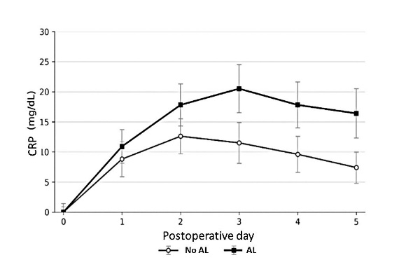
In a systematic review by Dost et al.15 that included 20 articles, the utility of CRP and PCT in the setting of AL was evaluated. The authors reported that, in patients with an uncomplicated postoperative course, CRP levels peak on POD 3 and subsequently decline. In contrast, levels remain elevated in patients who develop complications. Consistent with the findings of Bona et al.,14 the authors concluded that low CRP levels help rule out AL and may support discharge decisions in this patient population. Regarding PCT, they also emphasized the importance of low levels in this setting and suggested that PCT may have greater specificity than CRP.
The best results for detecting anastomotic complications are obtained by assessing multiple markers simultaneously. A retrospective study by Hu et al.¹⁶ demonstrated that PCT and CRP are valuable markers for detecting AL. On POD 3, CRP had a sensitivity of 96% and a specificity of 89.4%, whereas PCT had a sensitivity of 86% and a specificity of 93.5%. However, simultaneous measurement of both markers on POD 3 improved diagnostic accuracy for AL, yielding a specificity of 90% and a sensitivity of 100%.
In another retrospective series, PCT was the best predictor of AL on POD 3 compared with CRP and WBC count.17 On POD 5, a PCT cutoff value of 4.93 mg/mL yielded the highest sensitivity, specificity, and NPV. The predictive performance of PCT improved substantially when combined with CRP, WBC count, or both.
The costs associated with serum biomarker–guided protocols warrant careful consideration. Although routine biomarker monitoring incurs additional costs, these may be offset by potential savings resulting from the prevention of complications, shorter hospital stays, and more efficient resource utilization. The high NPV of certain biomarkers suggests that costs may be reduced by enabling early and safe discharge of low–risk patients. However, implementation of these protocols requires careful consideration of local resources, laboratory capacity, and the limitations of the healthcare system.
CONCEPTUAL SUMMARY
Serum biomarkers have assumed an increasingly important role in the early diagnosis of anastomotic leakage (AL), although heterogeneity across studies limits comparisons of results and the establishment of universal cutoff values. Their primary utility lies in complementing clinical assessment and facilitating early risk stratification to identify patients at low or high risk of complications.
Among the biomarkers studied, C–reactive protein (CRP) and procalcitonin (PCT) are supported by the strongest body of evidence. Both have a high negative predictive value, and low levels during the first few postoperative days can help rule out AL and support early hospital discharge. PCT may offer greater specificity than CRP, particularly when measured between postoperative days 3 and 5.
Current evidence suggests that no single biomarker is sufficient to diagnose AL. The best results are obtained by combining multiple biomarkers, particularly CRP and PCT, with clinical assessment. Accordingly, these biomarkers should be regarded as adjuncts to clinical evaluation that may facilitate early detection of complications and, particularly, identification of patients at very low risk of AL.
REFERENCES
1. Wright EC, Connolly P, Vella M, Moug S. Peritoneal fluid biomarkers in the detection of colorectal anastomotic leaks: a systematic review. Int J Colorectal Dis. 2017;32(7):935–45.
2. Ben–David M, Carmeli I, Orgad R, et al. Implantation of an Impedance Sensor for Early Detection of Gastrointestinal Anastomotic Leaks. J Surg Res. 2022;278:49–56.
3. Corke C, Glenister K. Monitoring intestinal ischaemia. Crit Care Resusc. 2001 Sep;3(3):176–80. PMID: 16573500.
4. Gray M, Marland JRK, Murray AF, et al. Predictive and Diagnostic Biomarkers of Anastomotic Leakage: A Precision Medicine Approach for Colorectal Cancer Patients. J Pers Med. 2021;11(6):471.
5. Yang L, Huang XE, Xu L, et al. Acidic pelvic drainage as a predictive factor for anastomotic leakage after surgery for patients with rectal cancer. Asian Pac J Cancer Prev. 2013;14(9):5441–7.
6. Yung HC, Daroch AK, Parikh R, et al. Diagnostic Modalities for Early Detection of Anastomotic Leak After Colorectal Surgery. J Surg Res 2023;301:520–33.
7. Singh PP, Zeng IS, Srinivasa S, et al. Systematic review and meta–analysis of use of serum C–reactive protein levels to predict anastomotic leak after colorectal surgery. Br J Surg. 2014;101(4):339–46.
8. Stephensen BD, Reid F, Shaikh S, et al. PREDICT Study Group collaborators. C–reactive protein trajectory to predict colorectal anastomotic leak: PREDICT Study. Br J Surg 2020;107(13):1832–7.
9. Ge X, Cao Y, Wang H, et al. Diagnostic accuracy of the postoperative ratio of C–reactive protein to albumin for complications after colorectal surgery. World J Surg Oncol. 2017;15(1):15.
10. Maruna P, Nedelníková K, Gürlich R. Physiology and genetics of procalcitonin. Physiol Res. 2000;49 Suppl 1:S57–61. PMID: 10984072.
11. Zawadzki M, Czarnecki R, Rzaca M, et al. C–reactive protein and procalcitonin predict anastomotic leaks following colorectal cancer resections – a prospective study. Wideochir Inne Tech Maloinwazyjne. 2016;10(4):567–73.
12. Zhang G, Lian R, Sun L, et al. Redefined hyponatremia as a marker to exclude the diagnosis of anastomotic leakage after colorectal cancer surgery. J Int Med Res. 2020;48(8):300060520950565.
13. Käser SA, Furler R, Evequoz DC, et al. Could hyponatremia be a marker of anastomotic leakage after colorectal surgery? A single center analysis of 1,106 patients over 5 years. Langenbeck’s Arch Surg. 2014;399:783–8.
14. Bona D, Danelli P, Sozzi A, et al. C–reactive Protein and Procalcitonin Levels to Predict Anastomotic Leak After Colorectal Surgery: Systematic Review and Meta–analysis. J Gastrointest Surg. 2023;27(1):166–79.
15. Dost W, Rasully MQ, Zaman MN, et al. Predictive Biomarkers for the Early Detection of Anastomotic Leaks in Colorectal Surgeries: A Systematic Review. Cureus. 2024;16(11):e74616.
16. Hu Y, Ren J, Lv Z, Liu H, Qiu X. Procalcitonin and C–reactive protein as early predictors in patients at high risk of colorectal anastomotic leakage. J Int Med Res. 2024;52(6):3000605241258160.
17. El Zaher HA, Ghareeb WM, Fouad AM, et al. Role of the triad of procalcitonin, C–reactive protein, and white blood cell count in the prediction of anastomotic leak following colorectal resections. World J Surg Oncol. 2022;20(1):33.
C) IMAGING STUDIES
Unfortunately, no imaging modality is entirely reliable for detecting AL after colorectal surgery. Contrast enema and ultrasonography are inferior to CT and have fallen largely out of use over the past several years.1 Water–soluble contrast enema has traditionally been used to assess the integrity of colorectal anastomoses. The technique relies on direct visualization of the bowel lumen and the potential extravasation of contrast into the extraluminal space, a finding that is highly specific for AL. Despite its high specificity, however, contrast enema has very low sensitivity for detecting AL, particularly in cases of small or contained leaks. This limitation can lead to false–negative results, restricting its usefulness as an initial diagnostic test in patients with suspected AL.¹ Another important limitation is its inability to adequately assess extraluminal structures. Consequently, this technique has largely been superseded by more effective imaging modalities.2
One setting in which contrast enema remains relevant is the evaluation of patients before reversal of a diverting ostomy. In these cases, the study can confirm the absence of persistent leaks, strictures, or fistulous tracts, providing useful information for surgical decision–making.
The utility of ultrasonography for diagnosing AL is limited. Its main advantages are widespread availability, low cost, lack of ionizing radiation, and the ability to perform serial examinations. These characteristics make it particularly useful in critically ill patients or those who cannot readily be transported. In this setting, ultrasonography may help identify intra–abdominal complications.3
Direct visualization of the anastomotic defect is virtually impossible with ultrasonography. Instead, the technique can detect associated findings, including free fluid, intra–abdominal collections, and inflammatory changes in adjacent tissues. Complex collections containing internal echoes or air bubbles may suggest localized sepsis and represent a potentially relevant finding in patients with suspected AL4 However, these findings are nonspecific and must be interpreted in conjunction with the patient's clinical course and other, more sensitive and specific imaging studies.
Another important role of ultrasonography is monitoring the evolution of fluid collections. Serial examinations allow clinicians to assess the progression or resolution of previously identified collections and evaluate the response to antibiotic therapy or percutaneous drainage. This capability is particularly useful in patients managed nonoperatively, in whom close follow–up is essential for detecting treatment failure or progression of the septic process.4
Ultrasonography also plays an important therapeutic role by guiding percutaneous drainage. In patients with accessible collections, it enables minimally invasive procedures with high success rates, helping achieve source control without reoperation. This approach is particularly beneficial in selected, hemodynamically stable patients with well–defined, readily accessible collections. However, CT remains the preferred modality for planning and guiding drainage of deep or difficult–to–access collections.4
Despite these advantages, ultrasonography has important limitations in this setting, including interference from bowel gas, obesity, the deep location of anastomoses, and the complex anatomy of the postoperative abdomen. In addition, ultrasonography is operator–dependent, introducing variability in the interpretation of findings. For these reasons, its sensitivity for the specific diagnosis of AL is substantially lower than that of CT, limiting its role as a definitive diagnostic test.5
CT is undoubtedly the most useful imaging modality for diagnosing postoperative complications after colorectal surgery, as it provides information about the anastomosis and surrounding tissues. However, false–negative results may occur in up to 25% of patients with AL. Therefore, particular caution is warranted to avoid delaying active intervention when CT findings are unremarkable.6 In the series reported by Doeksen et al.,7 the false–negative rate may have exceeded 50%, meaning that half of the patients who underwent reoperation had normal CT findings. It is therefore important to emphasize that the decision to reoperate should be based primarily on the clinical presentation rather than on the results of imaging studies.
Delaying reoperation in this setting increases hospital length of stay (LOS) and, particularly, mortality, which rises from 4% to 45%–62.5% among patients with false–negative CT findings and a consequent delay in reoperation.8 It is also important to note that CT sensitivity and specificity are lower during the first few postoperative days and increase as time progresses. Paradoxically, the period when an early decision to reoperate is most critical is often when CT is least sensitive. Therefore, when clinical suspicion of AL is high, an unremarkable CT scan should not delay reintervention. In these cases, clinical judgment takes precedence over imaging findings because delays in diagnosis and treatment can have serious consequences for the patient.
Intravenous contrast administration allows adequate assessment of tissue perfusion, the integrity of abdominal structures, and associated inflammatory changes, all of which are essential for detecting postoperative complications. Accordingly, intravenous contrast–enhanced CT is considered the standard approach because it significantly improves diagnostic performance compared with unenhanced CT.9
Intravenous contrast improves the detection of indirect signs of AL, including changes in the perianastomotic fat and, more importantly, collections with peripheral enhancement. The latter finding is particularly relevant because enhancement of the wall of a collection associated with an active infectious process suggests that percutaneous drainage may be necessary. In addition, assessment of contrast distribution helps distinguish simple free fluid from organized collections, with direct implications for therapeutic decision–making. Identifying the number, location, and size of collections, as well as their relationship to adjacent structures, is essential for determining whether percutaneous drainage is feasible and planning the appropriate approach. Intravenous contrast also facilitates the detection of associated complications, such as venous thrombosis, distant abscesses, or involvement of other organs, extending its diagnostic value beyond the specific assessment of AL.9
Another, more difficult aspect to assess with this technique is anastomotic perfusion. Areas of reduced perfusion or heterogeneous enhancement may suggest vascular compromise or ischemia, factors closely associated with the development and progression of AL. Thus, intravenous contrast contributes not only to the diagnosis of leakage but also to the identification of its potential causes, which can be valuable when planning a surgical strategy in patients requiring reintervention.10
Intravenous contrast–enhanced CT is an indispensable tool in the evaluation of suspected AL. Its main contribution lies in identifying indirect signs of leakage and detecting collections. Although it does not always permit direct visualization of contrast extravasation, its routine use significantly improves diagnostic accuracy and is essential for treatment planning.
There are absolute and relative contraindications to intravenous contrast–enhanced CT.11,12 The most relevant absolute contraindication is a history of a previous severe anaphylactic reaction to iodinated contrast material. Reexposure in these patients may trigger potentially fatal events; therefore, contrast administration should be avoided and alternative diagnostic modalities considered. Relative contraindications primarily include renal insufficiency, particularly in patients with substantial impairment of renal function. Although the risk of contrast–induced nephropathy is now considered lower than previously thought, it remains a consideration, especially in patients with advanced chronic kidney disease, diabetes mellitus, or dehydration. In these cases, the decision to administer contrast should be individualized by weighing the diagnostic benefit against the potential risk and implementing preventive measures, such as adequate hydration.
It is important to note that patients with AL may have impaired renal function related to postoperative ileus and subsequent dehydration. In patients with suspected AL, intravenous contrast–enhanced CT remains the preferred diagnostic study, even in those with impaired renal function. Current evidence suggests that the risk of acute kidney injury associated with intravenous iodinated contrast has been overestimated, with no significant increase in risk compared with unenhanced studies in most patients.13 Accordingly, no absolute contraindication is based solely on serum creatinine or estimated glomerular filtration rate (eGFR) values. An individualized assessment is recommended, particularly in patients with an eGFR below 30 mL/min. In critical clinical settings, the benefits of contrast administration generally outweigh the potential risks.14
Oral contrast is frequently administered, although its actual usefulness is currently debatable. Its use has been proposed to improve the detection of AL, particularly by directly demonstrating contrast extravasation. The theoretical benefit is increased diagnostic specificity through unequivocal visualization of a leak. In clinical practice, however, the value of oral contrast remains controversial. Studies have shown that oral contrast does not significantly increase CT sensitivity compared with intravenous contrast alone, particularly in cases of early or low–output leaks.15 This is partly because oral contrast may not reach the anastomosis by the time of imaging, particularly in patients with postoperative ileus, a common occurrence in this setting.
Another important consideration is that oral contrast administration may delay imaging, which is particularly problematic in patients with suspected abdominal sepsis, in whom time to diagnosis is a major determinant of clinical outcome.15 Therefore, prioritizing rapid, readily available studies, such as intravenous contrast–enhanced CT, is often a more appropriate strategy. Patients with AL frequently have ileus or cessation of intestinal transit, which reduces the likelihood that orally administered contrast will reach the anastomosis. This makes rectal contrast particularly relevant (Table 1). In selected cases, placement of a nasogastric tube may be useful in patients unable to tolerate oral intake, particularly those with postoperative ileus. In patients with AL, postoperative ileus is common, and its pathophysiology may involve both a functional component and partial mechanical obstruction secondary to inflammation or perianastomotic collections. In this setting, the osmotic effect of water–soluble contrast may help resolve associated partial obstructive symptoms.16
Table 1. Effects of rectal contrast. Adapted from Marres et al.15
With rectal contrast (n=50) | Without rectal contrast (n=175) | p | |
Contrast at the anastomosis | 26% | 81.7% | <0.001 |
False–negative CT scans | 18% | 4.6% | 0.004 |
Mortality | 16% | 4.6% | 0.006 |
Failure to rescue | 19.5% | 7.9% | 0.048 |
Oral contrast may also complicate image interpretation by producing artifacts or obscuring small extraluminal air bubbles, which are important indirect signs of AL. In addition, oral contrast may be poorly tolerated by patients with clinical deterioration, nausea, or an increased risk of aspiration.
Most current guidelines agree that intravenous contrast–enhanced CT is sufficient as the initial imaging study when AL is suspected, reserving oral contrast for selected situations.9 These may include subacute presentations, suspected contained leaks, or cases in which more precise characterization of communication between the bowel lumen and a collection is needed (Fig. 1).
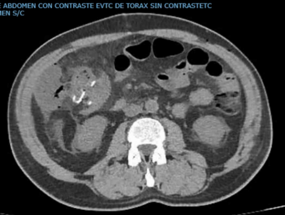
In summary, oral contrast has limited value in the diagnosis of AL and should not be used routinely. Its use should be individualized according to the clinical context, anastomotic location, and urgency of diagnosis, avoiding routine administration in acute settings where it may delay therapeutic decision–making.
The most important imaging finding is undoubtedly contrast extravasation. Acute contrast reactions are much more common with intravenous administration and are rarely observed after rectal administration. When contrast is administered rectally, it reaches the anastomosis in 81.7% of cases, compared with 26% when administered orally.15 Although the study included patients who underwent various types of resection and did not provide a subgroup analysis by surgical procedure, it is reasonable to expect that the more proximal the anastomosis, the lower the diagnostic utility of rectal contrast.
In 2021, Marres et al.15 analyzed 225 patients who underwent CT for suspected AL, of whom 175 (77.8%) received rectal contrast. The false–negative rate decreased from 18% to 4.6% when rectal contrast was administered. The authors also reported a difference in mortality between the two groups: 16% among patients who did not receive rectal contrast and 4.6% among those who did. This difference may be attributable primarily to delays in deciding to reoperate on patients with AL when CT findings are falsely negative. However, the literature is somewhat inconsistent regarding the sensitivity and specificity of rectal contrast–enhanced CT, largely because of differences in contrast administration techniques. The study cited above emphasized that 1 L of water–soluble contrast should be administered through a cannula positioned 40 cm above the patient's level, without inflating the balloon.
Huiberts et al.,17 from the Netherlands, analyzed a series of 108 CT examinations performed after colorectal surgery for suspected AL. Contrast extravasation was the only definitive CT finding of AL in their series. The authors therefore emphasized the importance of proper contrast administration to ensure adequate evaluation of the anastomosis. Their findings also suggest that interobserver variability is lower for the detection of contrast extravasation than for other findings, such as free fluid or perianastomotic air bubbles.
In 2023, Moreno–López et al.18 compared CT findings obtained with and without rectal contrast in patients with suspected AL, as interpreted by two independent observers. Agreement between the observers was high for the identification of contrast leakage on CT, demonstrating that this finding is unequivocal and readily recognized. In addition, the false–negative rate decreased for both observers when rectal contrast was administered, from 23% to 18% (p=0.02) and from 32% to 16% (p=0.0001).
Published evidence comparing the use of rectal contrast after right versus left colectomy is limited, but the diagnostic benefit of rectal contrast appears to be lower for ileocolic anastomoses. This difference in diagnostic performance may be attributable to the greater difficulty of obtaining adequate images in patients who have undergone right colectomy, particularly in ensuring that the administered intraluminal contrast reaches the anastomosis, which is generally farther from the point of contrast administration. In general, oral contrast is considered more useful for right–sided anastomoses, whereas rectal contrast is favored for left–sided anastomoses. However, as discussed above, oral contrast does not always reach ileocolic anastomoses adequately because of the frequent postoperative ileus associated with these presentations.
Unfortunately, rectal contrast administration in patients with suspected AL is not without risk. By increasing intraluminal pressure, it could potentially contribute to disruption of a recent anastomosis and bacterial dissemination into the pelvis or even through the bloodstream.19 These effects may be particularly relevant in the early postoperative period and in patients with distal anastomoses, where mechanical forces and the local bacterial burden are greater. Accordingly, the routine use of rectal contrast has been questioned. Although the risk of bacterial dissemination associated with rectal contrast has been described primarily in the context of contrast enema, this phenomenon is also theoretically possible during rectal contrast–enhanced CT. Nevertheless, its incidence is likely very low because smaller volumes are used and contrast is administered at low pressure, reducing colonic distension and the potential extravasation of contaminated contents.
Several CT findings may raise suspicion for AL (Figs. 2 and 3):
– Extraluminal contrast extravasation
– Free intraperitoneal fluid
– Perianastomotic air bubbles
– Pneumoperitoneum
The first three findings may be normal in the postoperative period, particularly during the first few days. Pneumoperitoneum may persist until postoperative day 10; however, its volume may raise concern for an anastomotic complication. Furthermore, an increase in pneumoperitoneum between CT scans performed on different postoperative days strongly suggests an anastomotic complication or a leak from another hollow viscus.
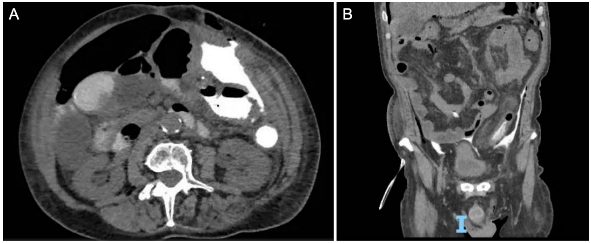
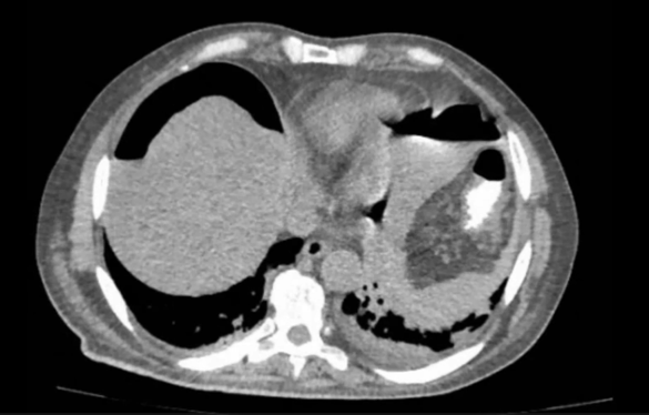
A similar principle applies to perianastomotic air bubbles and free intraperitoneal fluid. Both are common findings during the first few postoperative days, but a substantial amount of either should raise suspicion for AL. Gervaz et al.20 reported that the presence of more than 500 mL of intraperitoneal fluid suggests AL.
Comparing CT scans obtained on different postoperative days may provide an additional useful parameter. If the complication remains unresolved, an increase in free fluid or pneumoperitoneum would be expected on subsequent CT scans. Repeat CT examinations, however, should be considered only in hemodynamically stable patients without clinical deterioration.
Regarding septic complications in the pelvis, CT is less useful after LAR (Fig. 4). Magnetic resonance imaging (MRI) may therefore be an alternative in these cases because it provides important information about the pelvis and the anastomosis. For the detection of AL, MRI has a sensitivity of 94%, specificity of 80%, positive predictive value of 94%, and negative predictive value of 80%.21 MRI is particularly useful for evaluating local complications, such as abscesses, presacral sinuses, and complex fistulas, that may not be fully characterized by CT.
Nevertheless, MRI has important limitations. Limited availability in emergency settings restricts its use in the acute setting. In addition, surgeons may be less familiar with interpreting MRI findings than CT images and may depend on reports from radiologists. For these reasons, MRI is not generally included in the initial diagnostic algorithm for patients with suspected AL. In current practice, MRI is reserved for specific indications, primarily the detailed evaluation of complex pelvic complications, fistulas, or persistent collections, and as an adjunct to other imaging modalities when their findings are inconclusive.21
CONCEPTUAL SUMMARY
No imaging study is infallible for the diagnosis of AL. CT is currently the preferred imaging modality because it can identify indirect signs of leakage, including collections, free fluid, pneumoperitoneum, and inflammatory changes. However, its sensitivity is limited, and false–negative results may be common, particularly during the first few postoperative days.
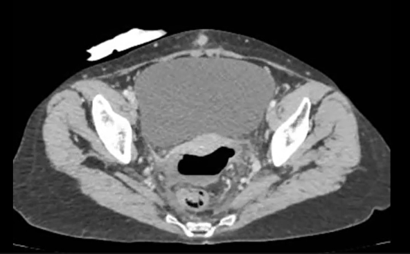
Therefore, a normal CT scan does not exclude AL when clinical suspicion is high, and the decision to reoperate should be based primarily on the patient's clinical course. Intravenous contrast administration is considered essential for optimizing the diagnostic performance of CT. Oral contrast offers limited benefits and may delay diagnosis; therefore, its routine use is not recommended. In contrast, rectal contrast has been shown to improve diagnostic sensitivity, particularly for colorectal anastomoses, by enabling direct visualization of contrast extravasation, the most specific CT sign of AL.
Ultimately, imaging studies are essential diagnostic tools, but their findings should never take precedence over the patient's clinical assessment.
REFERENCES
1. Chierici A, Granieri S, Frontali A. Diagnostic accuracy of water–soluble contrast enema, contrast–enema computed tomography and endoscopy in detecting anastomotic leakage after (Colo) proctectomy: A meta–analysis. Colorectal Dis. 2023;25(7):1371–1380.
2. Marres CCM, van de Ven AWH, Leijssen LGJ, et al. Colorectal anastomotic leak: delay in reintervention after false–negative computed tomography scan is a reason for concern. Tech Coloproctol. 2017;21(9):709–714.
3. Lin CM, Hung GU, Chao TH, et al. The limited use of ultrasound in the detection of abdominal abscesses in patients after colorectal surgery: compared with gallium scan and computed tomography. Hepatogastroenterology. 2005;52(61):79–81.
4. Hedrick TL, Kane W. Management of Acute Anastomotic Leaks. Clin Colon Rectal Surg. 2021;34(6):400–405.
5. Sartelli, M., Coccolini, F., Kluger, Y. et al. WSES/GAIS/SIS–E/WSIS/AAST global clinical pathways for patients with intra–abdominal infections. World J Emerg Surg 2021: 16, 49.
6. Gessler B, Eriksson O, Angenete E. Diagnosis, treatment, and consequences of anastomotic leakage in colorectal surgery. Int J Colorectal Dis. 2017;32:549–556.
7. Doeksen A, Tanis PJ, Wüst AF, et al. Radiological evaluation of colorectal anastomoses. Int J Colorectal Dis. 2008;23(9):863–8.
8. Kornmann VN, van Ramshorst B, Smits AB, et al. Beware of false–negative CT scan for anastomotic leakage after colonic surgery. Int J Colorectal Dis. 2014;29(4):445–51.
9. Hernandez PT, Paspulati RM, Shanmugan S. Diagnosis of Anastomotic Leak. Clin Colon Rectal Surg. 2021;34(6):391–399.
10. Kaur P, Karandikar SS, Roy–Choudhury S. Accuracy of multidetector CT in detecting anastomotic leaks following stapled left–sided colonic anastomosis. Clin Radiol. 2014;69(1):59–62.
11. Wang C, Ramsey A, Lang D, et al. Management and Prevention of Hypersensitivity Reactions to Radiocontrast Media: A Consensus Statement from the American College of Radiology and the American Academy of Allergy, Asthma & Immunology. Radiology. 2025;315(2):e240100.
12. Davenport MS, Perazella MA, Yee J, et al. Use of intravenous iodinated contrast media in patients with kidney disease: consensus statements from the American College of Radiology and the National Kidney Foundation. Radiology. 2020;294(3):660–668.
13. European Society of Urogenital Radiology. ESUR Guidelines on Contrast Agents. Version 10.0. 2018/2022.
14. Obed M, Gabriel MM, Dumann E, et al. Risk of acute kidney injury after contrast–enhanced computerized tomography: a systematic review and meta–analysis of 21 propensity score–matched cohort studies. Eur Radiol. 2022;32(12):8432–8442.
15. Marres CCM, Engelmann EWM, Buskens CJ, et al. The importance of rectal contrast in CT assessment to detect anastomotic leakage after colorectal surgery. Colorectal Dis 2021;23(9):2466–2471.
16. Catena F, Di Saverio S, Kelly MD, et al. Bologna Guidelines for Diagnosis and Management of Adhesive Small Bowel Obstruction (ASBO): 2010 Evidence–Based Guidelines of the World Society of Emergency Surgery. World J Emerg Surg. 2011;6:5.
17. Huiberts AA, Dijksman LM, Boer SA, et al. Contrast medium at the site of the anastomosis is crucial in detecting anastomotic leakage with CT imaging after colorectal surgery. Int J Colorectal Dis. 2015;30(6):843–8.
18. Moreno–Lopez N, Mvouama S, Bourredjem A, et al. CT scan for early diagnosis of anastomotic leak after colorectal surgery: is rectal contrast useful? Tech Coloproctol. 2023;27(8):639–645.
19. Yu, L., Chen, G., Wang, H. et al. MRI in diagnose post–operative anastomotic leak in patients with rectal cancer: preliminary experience. BMC Surg 2022;22:422
20. Gervaz P, Platon A, Buchs NC, et al. CT scan–based modelling of anastomotic leak risk after colorectal surgery. Colorectal Dis 2013;15(10):1295–300
21. de Wit A, Daams F. Considerations in case of suspected anastomotic leakage in the lower GI tract. Best Pract Res Clin Gastroenterol. 2024 Jun; 70:101925
VII. Treatment
The management of AL varies considerably depending on several factors, including the patient's overall condition, the type of anastomosis, and the extent of the defect.
As previously described, a widely used classification of AL is based on the therapeutic approach required for its management. Grade AL can be managed conservatively, without invasive intervention. Grade B AL requires percutaneous drainage (PD) or endoscopic treatment. Grade C AL is the most severe and requires surgical reintervention for definitive management.
GRADE A ANASTOMOTIC LEAKAGE – NONOPERATIVE MANAGEMENT
Nonoperative management may be initiated in a selected group of hemodynamically stable patients without signs of sepsis. The two mainstays of source control in this setting are bowel rest and antibiotic therapy.
Although bowel rest is commonly listed as a cornerstone of AL management, its theoretical rationale is less clear. No randomized controlled trials have been published comparing fasting with continued oral intake in patients with AL. Bowel rest is generally incorporated into broader treatment strategies, making it difficult to isolate its independent effect. Nevertheless, it is reasonable to hypothesize that reducing intestinal transit may decrease the bacterial load and the amount of fecal material passing through the leak site.
Bowel rest is also justified by the frequent occurrence of ileus, feeding intolerance, abdominal distension, nausea, vomiting, and overall clinical deterioration in patients with AL, all of which may make enteral nutrition impractical or unsafe. Under these circumstances, withholding oral intake may help prevent complications such as aspiration, worsening ileus, increased abdominal pain, and hemodynamic deterioration.1
In some cases, prolonged bowel rest necessitates total parenteral nutrition (TPN), which has become an important component of conservative management in selected patients.2
From a pathophysiological standpoint, AL is associated with a hypercatabolic state, a systemic inflammatory response, alterations in protein and energy metabolism, and substantial nutritional deterioration. These processes are exacerbated by abdominal sepsis, ileus, feeding intolerance, and the frequent need for bowel rest, which limits or precludes enteral nutrition. In this setting, TPN provides adequate macronutrient and micronutrient intake, helps preserve lean body mass, supports immune function, and promotes tissue healing.2
Evidence specifically addressing TPN in patients with colorectal AL remains limited and is derived predominantly from observational studies, case series, and retrospective analyses, precluding high–level evidence–based recommendations.
Clinically, TPN is indicated primarily when enteral nutrition is not feasible or is contraindicated, including in patients with prolonged ileus, functional or mechanical obstruction, high–output fistulas, hemodynamic instability, uncontrolled sepsis, severe feeding intolerance, a high risk of aspiration, or an AL that precludes safe nutritional intake. In these cases, TPN provides essential metabolic support to maintain nutritional status while the infectious process resolves and the patient's clinical condition stabilizes.2
However, TPN carries risks, including central venous catheter–related infections, metabolic complications such as hyperglycemia, hepatic dysfunction, and electrolyte disturbances, thrombotic events, and increased costs.3 These limitations have led to broad agreement in the literature that enteral nutrition should be preferred whenever gastrointestinal function is preserved and feeding is safe and tolerated, with TPN reserved for specific, well–justified indications.
In contemporary colorectal surgery and the management of abdominal sepsis, nutritional support is viewed not as an isolated intervention but as an integral component of a comprehensive nonoperative management strategy. This approach includes effective source control, antimicrobial therapy tailored to microbiological findings and local resistance patterns, hemodynamic stabilization, and adequate drainage by surgical, percutaneous, or endoscopic means when necessary.
The decision to initiate TPN should be individualized on the basis of a multifactorial assessment that considers clinical, metabolic, infectious, anatomical, and functional variables, as well as the response to source control. Early involvement of a multidisciplinary team comprising surgeons, infectious disease specialists, clinical nutrition specialists, and critical care physicians is essential to optimize outcomes.
In summary, although high–level evidence specifically addressing TPN in patients with AL is limited, the available literature supports its use as part of nonoperative management in selected patients, particularly when enteral nutrition is not feasible or is unsafe. TPN should be incorporated into a comprehensive therapeutic strategy aimed at controlling the source of infection, resolving sepsis, and restoring metabolic status, with management individualized and adjusted according to the patient's clinical course.
Candidates for nonoperative management include patients with small leaks associated with collections smaller than 3 cm, provided that hemodynamic instability is absent.1 A similar clinical scenario involves patients who develop colocutaneous fistulas through abdominal incisions, with fecal material draining through the wound.
Antibiotic regimens should provide coverage against gram–negative organisms and anaerobes. To characterize the bacterial pathogens involved in AL, the 2013 CIAOW worldwide observational study4 collected data from 1,898 patients at 68 medical institutions worldwide. The most frequently identified pathogens causing intra–abdominal infections were Escherichia coli (35.7%), Enterococcus spp. (12.9%), Klebsiella spp. (10.5%), Pseudomonas aeruginosa (5.1%), and Enterobacter spp. (4.1%). A Korean study5 identified 11 different pathogens, with an average of approximately two bacterial species per patient. Consistent with the CIAOW findings, E. coli and Enterococcus spp. (26.8% each) were the most frequently isolated organisms, followed by P. aeruginosa (12.2%). Notably, 12.1% of the isolates were multidrug–resistant organisms belonging to the ESKAPE group (Enterococcus faecium, Staphylococcus aureus, Klebsiella pneumoniae, Acinetobacter baumannii, P. aeruginosa, and Enterobacter spp.). Methicillin–resistant S. aureus (MRSA) was the most frequently identified organism within this group. Several factors are associated with multidrug–resistant organisms, including immunosuppression, corticosteroid use, prior exposure to broad–spectrum antibiotics, liver or lung disease, and a hospital stay exceeding 5 days. In addition, the number of reoperations is closely associated with an increased prevalence of multidrug–resistant organisms (Fig. 1).6 Fungal superinfection is another consideration in this setting and has been reported in approximately 10% of patients with postoperative intra–abdominal abscesses (IAAs). Candida spp. is the most frequently isolated organism. Predisposing factors are similar to those associated with bacterial resistance described above.7
The most commonly used antibiotic regimens are piperacillin–tazobactam and ceftriaxone plus metronidazole.7 Local microbiological profiles and institutional patterns of bacterial resistance should guide antimicrobial selection, with adjustments made as appropriate. However, few studies have evaluated antibiotic regimens and bacterial resistance patterns specifically in patients who develop AL after colorectal surgery. Consequently, microbiological cultures are obtained in only a limited proportion of patients with AL to guide targeted antibiotic therapy. This issue is discussed further in the section on percutaneous drainage.
Prolonged antibiotic courses have traditionally been used in this setting. However, prolonged treatment and inappropriate antibiotic use are major contributors to the development of multidrug resistance. Rational and appropriate antibiotic use is important both for resolving the septic process and for reducing the likelihood of selecting for multidrug–resistant pathogens. The optimal duration of antibiotic therapy remains controversial. A multicenter study published in 2015 demonstrated that 4 days of intravenous antibiotic therapy were sufficient to treat intra–abdominal abscesses. Resolution of the septic process was similar with 4–day treatment and with longer courses.8
A French study published in 2018 evaluated antibiotic duration in critically ill patients admitted to the intensive care unit (ICU).9 The randomized DURAPOP trial included 249 patients with postoperative intra–abdominal infections, most of which occurred after colorectal surgery. All patients received intravenous antibiotics through day 8, at which point they were randomized either to discontinue antibiotics or to continue treatment for an additional 7 days (through day 15). No differences were observed between the groups in the need for reoperation, mortality, ICU length of stay, or hospital length of stay. However, there was a greater trend toward the need for percutaneous drainage among patients who discontinued antibiotic therapy on day 8. Nevertheless, an 8–day course of antibiotic therapy appears adequate for patients admitted to the ICU after colorectal surgery who develop AL Any change in the patient's clinical condition, or persistence of symptoms despite appropriate treatment, warrants repeat imaging because additional intervention may be required.
Special consideration should be given to patients with low anastomoses and a diverting ostomy who develop postoperative AL. In this setting, more than 80% of patients have a favorable outcome with conservative management.10 Factors associated with treatment failure include large, multiloculated abscesses, fistulas, and elevated Acute Physiology and Chronic Health Evaluation II (APACHE II) scores. Surgical management in this setting, involving takedown of the anastomosis, creation of an end ostomy, and closure of the rectal stump, carries a very high likelihood of a permanent ostomy, potentially exceeding 70%. In the absence of ischemia at the anastomotic ends, conservative management should be pursued.
In 2025, the first systematic review of the management of AL following rectal cancer surgery was published.10
Among the 7,236 patients included in the review, 850 developed AL. The success rate of nonoperative management was 44%. Short–term mortality was 0%; however, 55.6% of patients required subsequent intervention. In this setting, percutaneous drainage is an option when initial nonoperative management fails.
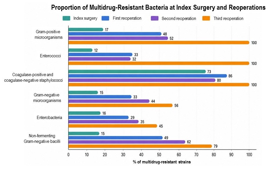
CONCEPTUAL SUMMARY
Nonoperative management of AL is appropriate for selected patients who are hemodynamically stable and have no signs of severe sepsis. The mainstays of treatment are appropriate antimicrobial therapy to control infection and supportive care, including bowel rest and nutritional support as needed.
Antimicrobial therapy should cover Gram–negative bacilli and anaerobes, with consideration given to the increasing prevalence of multidrug–resistant organisms and the possibility of fungal superinfection in selected patients. The optimal duration of antimicrobial therapy remains controversial, although current evidence suggests that shorter courses may be sufficient when adequate source control has been achieved.
Close clinical monitoring is essential, and repeat imaging is warranted if symptoms persist or worsen.
GRADE B ANASTOMOTIC LEAKAGE – PERCUTANEOUS MANAGEMENT
Intra–abdominal abscesses (IAAs) are relatively common after abdominal surgery and can substantially increase morbidity and mortality if not adequately treated. They are defined as collections of purulent fluid within the abdominal cavity, generally resulting from intraoperative contamination and/or AL. The incidence varies according to the type of index operation, the patient's overall condition, and intraoperative factors, and may reach 10%.11
In recent years, two terms have been introduced in the management of AL: passive and active drainage. Passive drainage refers to drainage without active suction and includes drains placed under ultrasound, CT, or endoscopic guidance. Active drainage involves a catheter connected to a suction system. The following section focuses on passive drainage.
In some cases of AL, particularly after colorectal anastomoses, the local inflammatory response, omentum, and adjacent structures can contain the spread of luminal contents, resulting in a perianastomotic collection.11
Risk factors for the development of IAAs include inflammatory bowel disease, emergency surgery, malnutrition, corticosteroid therapy, intraoperative contamination, preoperative RT, and splenectomy.11
Patients undergoing splenectomy have a threefold higher risk of developing postoperative collections. This may be attributable to the frequent occurrence of hematomas in this setting, together with an impaired immune response.12
In recent years, the management of AL has undergone a paradigm shift, driven by advances in diagnostic imaging and the development of minimally invasive techniques, which have enabled more conservative treatment approaches.
In this context, image–guided PD, performed under ultrasound or CT guidance, has emerged as a key tool for managing perianastomotic collections in selected patients.11
The primary goal is effective source control. PD should not be regarded as a lesser or merely temporary alternative to surgery; in many cases, it can provide definitive management of AL while preserving the anastomosis whenever possible. This approach has helped reduce reoperation rates, thereby decreasing associated morbidity and mortality and improving functional outcomes.
Persistence of an infected collection increases local pressure and reduces tissue perfusion, thereby impairing anastomotic healing. By evacuating purulent material and reducing the bacterial burden, PD may create a more favorable local environment for anastomotic preservation and better long–term function.
This approach is primarily used for grade B AL, characterized by a localized, contained collection in the absence of peritonitis or hemodynamic instability.13
Appropriate patient selection is fundamental to the success of PD. Inadequate patient selection is a major cause of treatment failure and delays in definitive management, with a negative impact on clinical outcomes.
The indications for PD are as follows:11
– Grade B AL.
– One or more perianastomotic collections with well–defined walls, identifiable on CT.
– Absence of generalized peritonitis.
– Clinical stability without hemodynamic compromise.
– Ability to ensure adequate clinical and imaging F-U.
Most IAAs are diagnosed relatively late, between postoperative days 5 and 10. In cases of very early AL, particularly within 48–72 hours after surgery, which often reflects technical failure or anastomotic ischemia, PD is less likely to be effective. The presence of a proximal ostomy increases the likelihood that PD will provide definitive treatment.14
Although the published literature on IAAs associated with AL is limited, most studies extrapolate management strategies from those used for pericolic collections associated with complicated diverticulitis, despite the substantial differences between these two clinical settings. In this context, collections larger than 3 cm are generally considered to warrant PD.14
Once an IAA has been diagnosed on CT, the collection must be assessed for suitability for PD. Close communication between the attending surgeon and the interventional radiology team is essential, as the timing and approach must be agreed upon by both teams. This process is dynamic and should be adapted to the patient's clinical condition and subsequent course. Planning begins with a detailed review of the CT images to assess the location and size of the IAA and its relationship to adjacent vascular and visceral structures.
The interventional radiology team plays a critical role in selecting the safest access route while minimizing the risk of injury to adjacent structures. The team must also assess whether catheter irrigation, exchange, or repositioning may be necessary.
The procedure is usually performed under local anesthesia with sedation, although general anesthesia may be required in critically ill patients. After needle puncture, correct catheter placement is confirmed by aspiration of fluid from the IAA, which is then sent for culture. International infectious disease guidelines recommend routinely obtaining cultures of the drained material, as the results may prompt a change in antimicrobial therapy when resistant organisms are identified.7 A catheter of appropriate caliber is then placed, typically ranging from 8 to 14 Fr, with larger catheters reserved for collections containing highly viscous material or fecal matter.14
The choice between ultrasound and CT guidance depends on several factors. Contrast–enhanced CT is the preferred imaging modality. However, ultrasound–guided PD may be performed in selected circumstances, such as when the collection is superficial and the operator has appropriate experience.11
The location of the IAA largely determines the access route and the likelihood of successful drainage. Intra–abdominal collections generally arise from proximal or intraperitoneal anastomoses. Drainage may be more challenging when small–bowel loops or vascular structures obstruct the access route. For pelvic collections associated with extraperitoneal anastomoses, drainage may be performed through a transgluteal, transvaginal, or transanal approach. The transgluteal approach is associated with greater morbidity, including fistula formation, gluteal abscesses, and pseudoaneurysms. Pain at the drainage site is the most common adverse event in patients undergoing transgluteal drainage. In selected circumstances, particularly in patients with a proximal ostomy, a transanastomotic approach may be useful. A drain is placed under direct visualization in the operating room. In the long term, anastomotic strictures or fecal incontinence may develop.13,15
PD does not mark the end of treatment; rather, catheter placement is only the first step. Subsequent management is critical to therapeutic success.
Close clinical monitoring is essential, including assessment of the patient's overall condition and drain output. Serial CT scans allow evaluation of collection size and detection of complications. In some cases, catheter exchange, placement of additional drains, or irrigation with normal saline may be necessary.
The duration of PD varies. Catheter removal is generally considered when output is minimal, the drainage fluid is serous, and imaging shows no residual collection. Despite appropriate patient selection, PD may fail. Failure is suggested by persistent or worsening signs of sepsis, lack of reduction in the IAA, or progressive clinical deterioration. In these cases, surgical intervention should not be delayed, as delays may adversely affect patient outcomes.
Rare cases of infection tracking along the drainage tract have been described, potentially leading to severe sepsis and extensive injury to surrounding tissues, including tissue necrosis. In this setting, immediate debridement is required to prevent further progression.
When drain output consists of fecal material or CT demonstrates clear contrast extravasation, a proximal ostomy is likely to be required to manage the complication.
CONCEPTUAL SUMMARY
PD is the treatment of choice for patients with grade B AL, characterized by a contained perianastomotic collection in the absence of generalized peritonitis or hemodynamic instability. The primary goal is effective source control using a minimally invasive approach, thereby allowing preservation of the anastomosis and avoidance of reoperation in many cases.
Appropriate indications and careful patient selection are critical to procedural success. PD is generally recommended for well–circumscribed collections larger than 3 cm that are accessible under CT or ultrasound guidance. The procedure also allows for the collection of specimens for microbiological culture to guide antibiotic therapy.
Following drain placement, close clinical and radiologic monitoring is essential to assess resolution of the collection and detect potential complications or treatment failure at an early stage. Persistent sepsis, failure of the collection to decrease in size, or clinical deterioration warrants reassessment of the management strategy and may ultimately necessitate surgical intervention.
GRADE B ANASTOMOTIC LEAKAGE – ENDOSCOPIC MANAGEMENT
Endoscopic management is indicated in hemodynamically stable patients with small anastomotic defects.
This approach is contraindicated in cases of significant tissue necrosis, endoscopic exposure of the peritoneum or bowel loops, frank communication with the abdominal cavity, or proximity of the defect to major blood vessels. Nevertheless, endoscopic techniques have gained increasing prominence in recent years for the management of colorectal AL.16
Main Strategies
Endo–VAC (Endoluminal Vacuum Therapy)
Negative–pressure wound therapy was first described for wound management in the 1990s. In 2001, Weidenhagen and colleagues introduced this technique in Munich, Germany, for the treatment of AL. Their initial report, published in 2008, described 29 patients treated with Endo–VAC and reported a 97% success rate.17
Since then, several commercial systems have become available in different countries, including Endosponge® (Braun Medical, Melsungen, Germany, 2022) and CNP (Lohmann & Rauscher, 2024). However, in most cases, the system is assembled using readily available materials, keeping costs relatively low. It can be constructed using a multifenestrated nasogastric tube and porous polyurethane foam, secured at the fenestrated end with nonabsorbable suture.
Most published studies originate from European countries, as the US Food and Drug Administration (FDA) has approved negative–pressure wound therapy systems for the treatment of wounds, ulcers, and burns but has not formally approved their use as endoluminal therapy. In US clinical practice, physicians may use devices and techniques approved for other indications when they consider them potentially beneficial to patients, provided that scientific evidence supports their use and informed consent is obtained. Accordingly, Endo–VAC is used off–label because it has not received specific FDA approval for this indication.
Endo–VAC is the most widely used endoscopic therapy for AL and is indicated in patients with extraperitoneal leaks, which are generally associated with perianastomotic or presacral collections. It has been used to treat anastomotic defects involving up to 75% of the anastomotic circumference, provided that the bowel ends remain viable, as well as associated cavities measuring up to 20 cm in diameter.18
The mechanism of action relies on continuous suction, which reduces bacterial contamination, local edema, and secretion while improving tissue perfusion and promoting granulation tissue formation.16
The procedure is straightforward. First, endoscopy is performed to identify the defect and the associated cavity. Any material within the cavity is aspirated during the same procedure. Once intraperitoneal communication has been ruled out, the cavity is irrigated with saline or povidone–iodine solution. The size of the defect is then assessed, and a foam sponge is fashioned to match its dimensions. The endoscope is subsequently advanced through the anastomotic defect into the cavity. A tube is positioned at the deepest part of the cavity. The tube carrying the sponge is then advanced through the initially positioned larger–diameter tube, which is simultaneously withdrawn. Endoscopic visualization allows confirmation of proper sponge placement within the cavity. Finally, the multifenestrated tube is connected to a suction system, generating continuous negative pressure ranging from −75 to −125 mm Hg. In selected cases involving large defects, two sponges connected to independent vacuum systems may be placed. For distal defects, the vacuum system can be placed directly using digital guidance.16,17
Sponges are generally exchanged every 48 to 72 hours. Before removal, saline instillation through the tube is recommended to reduce pain. The procedure is repeated until the cavity becomes too small to accommodate another sponge or has decreased in size by 90% relative to its initial dimensions.
The evidence supporting Endo–VAC therapy remains limited, with substantial heterogeneity across studies in patient selection, definitions of treatment success, and systems used. In 2022, a systematic review and meta–analysis addressed this topic, including 24 studies and a total of 690 patients who underwent negative–pressure therapy for AL. The authors had also contributed to the original report published in 2008.18
The analyzed population included patients with AL following colorectal anastomosis for neoplastic, diverticular, or traumatic disease, as well as patients with rectal stump dehiscence after Hartmann procedure.
Among patients who had received neoadjuvant therapy, time to healing was longer (31.6 vs 12.3 days). RT impairs granulation and subsequent wound healing through downregulation of growth factors. Other risk factors for treatment failure include male sex, multivisceral resection, and reoperation after the initial resection. Early initiation of treatment is important: the success rate was 72.4% when therapy was initiated within the first 15 days, compared with only 27.8% when treatment began later (p=0.003).
Initially, a diverting proximal ostomy was considered mandatory for negative–pressure therapy because it simplifies treatment and prevents fecal contamination. However, in the aforementioned systematic review, only 76% of patients had a proximal ostomy. Thus, diversion of the leaking anastomosis with a proximal ostomy may not be mandatory for negative–pressure therapy, although it may improve the success rate. When the sponge can be positioned entirely outside the bowel lumen, fecal passage may be preserved. Conversely, if the sponge partially or completely occludes the intestinal lumen, treatment is unlikely to succeed, and the sponge may become displaced, compromising its effectiveness.
Kuhn et al.19 evaluated 20 patients with extraperitoneal AL who did not have a diverting ostomy. Although the cohort was small, it seems reasonable that negative–pressure therapy may be effective in selected circumstances, particularly when the sponge can be positioned entirely within the cavity, leaving only the suction tube within the bowel lumen. Only 2 patients in the cohort, both of whom had an unfavorable clinical course, required proximal diversion. However, long–term outcomes did not differ between the 2 groups, supporting the hypothesis that proximal diversion may be avoided in selected cases. Intestinal continuity was restored in 63% of patients following treatment, compared with reported restoration rates of approximately 40% after Hartmann procedure reversal.20 Complications occurred in 12.1% of patients. The most common was anastomotic stricture, which was successfully managed with balloon dilation. Less frequent complications included bleeding, which was generally mild and amenable to endoscopic management, pain, and displacement of the sponge into the abdominal cavity.
Long–term outcomes remain incompletely characterized, although results from the French GRECCAR group published in 2020 were encouraging.21An assessment of LARS scores was performed 6 months after treatment completion. Only 53% of patients had mild LARS symptoms, and only 3 of 47 patients required anastomotic dilation.
One of the main concerns with this therapy is its duration. In the systematic review, a mean of 6.8 sponges was used per patient, and the mean treatment duration was 23.4 days. When treatment extended over a longer period, patients were managed as outpatients. The proportion of patients treated in the outpatient setting may reach 40%, a substantial figure that can help reduce treatment costs.
Treatment failure is defined by local or systemic deterioration and may, in most cases, result from inadequate granulation tissue formation with persistent sepsis. Despite differences in the definitions of treatment success across studies, the overall success rate was 81.4%. These findings support negative–pressure therapy as a valid treatment option for AL in patients with low colorectal anastomoses.
In recent years, some evidence has emerged supporting the addition of early surgical closure (ESC) of the anastomotic defect, which may shorten treatment duration and improve functional outcomes. The negative–pressure therapy procedure is performed as described above. Once the cavity is covered with granulation tissue, ESC is performed. The procedure takes place in the operating room under general anesthesia, with the patient in the Lloyd–Davies position. Depending on the height of the defect, an anal retractor (eg, Lone Star®, CooperSurgical, Trumbull, CT) or a transanal minimally invasive surgery platform (eg, GelPOINT Path®, Applied Medical, Rancho Santa Margarita, CA) may be used. A drain is also placed and brought out a few centimeters distal to the anastomotic defect, through the rectal mucosa.
A systematic review published in 2022 included patients who underwent endoscopic vacuum therapy with or without ESC.22 Although no prospective randomized trials have been published to date, comparison of the 2 approaches showed that the addition of ESC reduced the mean number of sponges used by 6, directly affecting treatment costs and duration. ESC may also be associated with better functional outcomes and a lower rate of permanent ostomy. However, selection bias must be considered because patients with anastomotic leakage involving an ileal pouch were overrepresented in the ESC group. Permanent ostomy rates were 33% without ESC and 18% with ESC.
In the same year, the group led by Roel Hompes23 published its experience with negative–pressure therapy combined with surgical closure in patients who had undergone anterior resection for rectal cancer. After AL was diagnosed, laparoscopy and ileostomy creation were performed in patients who did not already have a diverting ileostomy from the initial operation. The study included 68 patients. Treatment was initiated within the first 21 days after the initial surgery in 50% of patients, and the proportion was similar between patients undergoing surgical closure with an anal retractor and those treated using a minimally invasive surgery platform.
Notably, a 100% success rate and satisfactory subsequent function were achieved in patients who had the following characteristics: a diverting ostomy at the initial operation, early diagnosis of AL by contrast–enhanced CT, use of a transanal platform, and treatment initiation within the first 14 postoperative days. In contrast, patients who had received neoadjuvant therapy developed larger abscesses and required longer treatment and more sponges.
A few isolated reports have examined the role of this endoscopic treatment in patients with ileal pouch–anal anastomotic leakage. In 2019, the Dutch group published long–term results comparing 3 groups: patients with AL managed conservatively (2002–2009, with antibiotics and PD), patients treated with Endo–VAC and ESC (2010–2017), and a control group of patients without anastomotic complications. Pouch function and pouch failure rates were similar between the control group and patients treated with Endo–VAC and ESC, whereas outcomes were substantially worse in patients managed conservatively. These findings highlight that early, active treatment, in this case with Endo–VAC and ESC, may allow patients to maintain long–term pouch function without impairment.
Clips and Suturing Devices
Suturing devices and clips designed for closure of full–thickness defects can also be used to treat ALs. Their main limitation is that they close the defect without addressing any associated extraluminal fluid collections. Therefore, their use requires confirmation that no undrained extraluminal collections are present.16 These techniques have consequently been recommended for selected ALs associated with fistulas (eg, rectovaginal or rectovesical fistulas) or for very small defects. Because anastomotic defects in the gastrointestinal tract are frequently contaminated, closure alone may be insufficient for successful treatment, as it generally fails in the presence of an ongoing infection. Accordingly, these techniques should be reserved for carefully selected patients.
The over–the–scope clip (OTSC) system (Ovesco Endoscopy AG, Tübingen, Germany) was introduced in 2007. Made of nitinol, these clips are available in different designs and sizes. Compared with conventional endoscopic clips, they can capture larger tissue volumes, including the full thickness of the bowel wall, while preserving tissue perfusion. This design is intended to minimize tissue necrosis and promote healing. Their use is generally limited to defects smaller than 15 mm and involving less than 25% of the anastomotic circumference. Placement requires suction of the target tissue into the cap; consequently, the system is less suitable for chronic ALs, in which tissue fibrosis and loss of elasticity may prevent adequate tissue capture.
Most of the published evidence consists of case reports and small case series. A 2020 systematic review included 3,025 patients from 85 studies who underwent OTSC placement for various indications, including gastrointestinal bleeding, perforations, fistulas, ALs, and closure of defects after endoscopic procedures.24 The population was heterogeneous, and only 29 patients had AL after colorectal surgery. The overall success rate was 75.9%, with technical and clinical success rates of 86% and 74%, respectively. These results should be interpreted in the context of the heterogeneous population, which also included patients with leaks involving the upper gastrointestinal tract.
A 2022 systematic review included 107 patients with colorectal ALs managed with OTSC.25 Approximately 50% required additional interventions, including percutaneous drainage and/or surgery. Technical and clinical success rates were 84% and 74%, respectively, similar to those reported in the earlier review.
OTSC should be limited to carefully selected cases, particularly defects smaller than 15 mm treated within the first 3 days after diagnosis. For chronic fistulas between organs, the time elapsed since fistula development may be less relevant, and OTSC placement may be considered at a later stage.25
The OverStitch system (Apollo Endosurgery, Austin, TX, USA) is a disposable, single–use device that requires a double–channel endoscope. It incorporates a needle that allows both continuous and interrupted suturing. Its use in the colon is limited by the narrow lumen and acute angulations. Most clinical experience has been gained in the stomach, whereas reports of its use in the colon and rectum remain limited to case reports and small series. The system may be effective for defects smaller than 2 cm when diagnosed early. Treatment of the associated extraluminal collection is generally also required.
Newer devices, including SutuArt (Apollo, Austin, TX, USA) and the X–Tack system (Boston Scientific, Marlborough, MA, USA), have been introduced in recent years, but their role in the management of ALs remains unclear.
Conventional clips may be useful for closing defects smaller than 1 cm. However, their effectiveness is limited by tissue fibrosis, which may interfere with proper clip placement and tissue approximation.
Stents
Stents are available in different materials, including metal, biodegradable polymers, and plastic, and with varying degrees of coverage (fully covered, partially covered, or uncovered). Self–expanding stents have become widely used for colonic obstruction, both for palliation and as a bridge to surgery. In selected cases, they may also be used to treat ALs. The goal is to cover the anastomotic defect and thereby prevent communication between the bowel lumen and the extraluminal space. However, this approach has not been widely adopted. The largest published case series to date, reported by Lamazza et al.,26 included 22 patients who underwent anterior resection (19 with extraperitoneal anastomoses and 3 with intraperitoneal anastomoses). The anastomotic defect involved 30% of the circumference and was confirmed by contrast enema. A rectovaginal fistula was identified in 10 patients, and 68% had a proximal stoma. The stent remained in place for a median period of 45 (range, 14–92) days, after which it was spontaneously expelled. The overall success rate was 84%. Although the rectovaginal fistulas did not close completely in 3 patients, the reduction in defect size facilitated subsequent surgical treatment.
The most common complications include stent migration, which may occur in up to 50% of cases, and extraluminal collections, which frequently require additional surgical or PD. Another important limitation is that the distal end of the stent must remain more than 4 cm from the anal verge to avoid tenesmus.
An alternative approach is the VAC–Stent (Microtech Europe GmbH, Düsseldorf, Germany), a 25–mm device that combines a stent with a negative–pressure system. Initially used for esophagogastric conditions, it has more recently been applied to colorectal ALs. The device consists of a nitinol stent covered with a polyurethane sponge connected to a continuous suction system operating at −80 to −120 mmHg. By covering the anastomotic defect while simultaneously draining perianastomotic collections, the VAC–Stent may combine the advantages of stent placement and endoluminal negative–pressure therapy. A potential advantage of this approach is that it may obviate the need for a proximal diverting stoma.
The VAC–Stent is generally left in place for 3 to 7 days. Before removal, instillation of 40 mL of saline is recommended to facilitate extraction. Suction should be discontinued 2 to 4 hours before removal.
Despite these theoretical advantages, clinical evidence remains limited. The largest published case series to date was reported by Heiss et al.²⁷ from Cologne, Germany, and included 12 VAC–Stent placements. The cohort was heterogeneous and included patients with a diverting stoma created at the index operation, patients without a stoma who subsequently developed ALs, and patients who underwent intraoperative VAC–Stent placement during the index procedure. In the latter group, prophylactic VAC–Stent placement may be viewed as a potential alternative to a diverting stoma. Among the 6 patients treated with a VAC–Stent without a proximal stoma, a mean of 3 stents per patient was required, and the mean hospital stay was 17 days. A 12–mm VAC–Stent was used in 5 cases, with migration occurring in 2. No migration was reported with the 25–mm device. Although the economic implications of this approach are considerable, the potential savings associated with stoma care and subsequent surgery to restore intestinal continuity may offset some of these costs. More rigorous studies are clearly needed to define the role of this technique in the management of patients with ALs.
CONCEPTUAL SUMMARY
Endoscopic treatment of ALs is indicated in hemodynamically stable patients with small defects and no extensive tissue necrosis, peritoneal exposure, or direct communication with the abdominal cavity. In recent years, it has emerged as a minimally invasive alternative that may preserve the anastomosis and avoid reoperation. Endo–VAC is the most widely used approach, particularly for extraperitoneal ALs associated with presacral or perianastomotic collections. Its mechanism of action combines continuous drainage, reduction of the bacterial burden, and promotion of tissue granulation, with reported success rates approaching 80%. Early treatment initiation and the presence of a proximal diverting stoma are associated with better outcomes.
Recently, available evidence has begun to support ESC of the anastomotic defect as a complementary strategy, suggesting potential benefits in reducing treatment duration while preserving functional outcomes.
Other options include endoscopic clips, suturing devices, and self–expanding stents, which are reserved for small defects or highly specific clinical scenarios. Although initial results are promising, the evidence supporting these approaches remains more limited than that supporting Endo–VAC. Their use should be tailored to the anatomic characteristics of the defect and the patient's clinical status.
REFERENCES
1. Hedrick TL, Kane W. Management of acute anastomotic leaks. Clin Colon Rectal Surg. 2021;34(6):400–5.
2. Weimann A, Braga M, Carli F, et al. ESPEN practical guideline: Clinical nutrition in surgery. Clin Nutr. 2021;40(7):4745–61.
3. Pittiruti M, Hamilton H, Biffi R, et al. ESPEN Guidelines on Parenteral Nutrition: central venous catheters (access, care, diagnosis and therapy of complications). Clin Nutr. 2009;28(4):365–77.
4. Sartelli M, Catena F, Ansaloni L, et al. Complicated intra–abdominal infections worldwide: the definitive data of the CIAOW Study. World J Emerg Surg. 2014;9:37.
5. Yang G, Kim CW, Lee SH. Patterns of antibiotics and pathogens for anastomotic leakage after colorectal cancer surgery. Korean J Clin Oncol. 2019;15(2):79–85.
6. Bassetti M, Eckmann C, Giacobbe DR, et al. Post–operative abdominal infections: epidemiology, operational definitions, and outcomes. Int Care Med. 2020;46(2):163–72.
7. Bonomo RA, Chow AW, Edwards MS, et al. 2024 Clinical Practice Guideline Update by the Infectious Diseases Society of America on Complicated Intra–abdominal Infections: Risk Assessment, Diagnostic Imaging, and Microbiological Evaluation in adults, children, and pregnant people. Clin Infect Dis 2024;79(Suppl 3):S81–S87.
8. Sawyer RG, Claridge JA, Nathens AB, et al. STOP–IT Trial Investigators. Trial of short–course antimicrobial therapy for intraabdominal infection. N Engl J Med. 2015;372(21):1996–2005.
9. Montravers P, Tubach F, Lescot T, et al. DURAPOP Trial Group. Short–course antibiotic therapy for critically ill patients treated for postoperative intra–abdominal infection: the DURAPOP randomised clinical trial. Int Care Med. 2018;44(3):300–10.
10. Walsh R, Murphy E, Ryan ÉJ, Cahill RA. Management of anastomotic leak after elective anterior resection for rectal cancer: A systematic review and pooled analysis. Colorectal Dis. 2025;27(12):e70335.
11. Taha M, Abouelsadat MK, Elfakharany M, et al. Postoperative Intra–abdominal abscess following general surgery: a systematic review of risk factors, prevention, and the role of laparotomy. Cureus. 2025;17(8):e91100.
12. Barmparas G, Lamb AW, Lee D, et al. Postoperative infection risk after splenectomy. Int J Surg 2015;18:105–9.
13. Baig MK, Zhao RH, Batista O, Uriburu JP, et al. Percutaneous postoperative intra–abdominal abscess drainage after elective colorectal surgery. Tech Coloproctol 2002;6(3):159–64.
14. Pastier C, Ben Hmida W, Lefèvre JH, et al. Management of low colorectal/coloanal anastomotic leak: results of a French National Intergroups Practice Survey (FRENCH–GRECCAR–SFCD). World J Surg. 2025;49(7):1747–56.
15. Keller DS, Talboom K, van Helsdingen CPM, Hompes R. Treatment modalities for anastomotic leakage in rectal cancer surgery. Clin Colon Rectal Surg. 2021;34(6):431–438.
16. Forcignanò E, Verra M, Lo Secco G, Arezzo A. Endoscopic management of anastomotic insufficiencies in the lower gi tract. Visc Med. 2025.
17. Weidenhagen R, Gruetzner KU, Wiecken T, et al. Endoscopic vacuum–assisted closure of anastomotic leakage following anterior resection of the rectum: a new method. Surg Endosc. 2008;22(8):1818–25.
18. Bemelman WA, Arezzo A, Banasiewicz T, Brady R, Espín–Basany E, Faiz O, Jimenez–Rodriguez RM. Use of sponge–assisted endoluminal vacuum therapy for the treatment of colorectal anastomotic leaks: expert panel consensus. BJS Open. 2022;6(5):zrac123.
19. Kühn F, Schardey J, Wirth U, et al. Endoscopic vacuum therapy for the treatment of colorectal leaks – a systematic review and meta–analysis. Int J Colorectal Dis. 2022;37(2):283–92.
20. Kühn F, Hasenhütl SM, Hofmann FO, et al. Endoscopic vacuum therapy for left–sided colorectal anastomotic leak without fecal diversion. Dis Colon Rectum. 2022;65(3):421–8.
21. Abdalla S, Cotte E, Epin A, et al. On behalf the French GRECCAR group. Short–term and Long–term Outcome of endoluminal vacuum therapy for colorectal or coloanal anastomotic leakage: results of a nationwide multicenter cohort study from the French GRECCAR Group. Dis Colon Rectum. 2020;63(3):371–80.
22. de Lacy FB, Talboom K, Roodbeen SX, et al. Endoscopic vacuum therapy and early surgical closure after pelvic anastomotic leak: meta–analysis of bowel continuity rates. Br J Surg. 2022;109(9):822–31.
23. Wasmann KA, Reijntjes MA, Stellingwerf M, et al. Endo–sponge assisted early surgical closure of ileal pouch–anal anastomotic leakage preserves long–term function: a cohort study. J Crohns Colitis. 2019;13(12):1537–45.
24. Bartell N, Bittner K, Kaul V. et al. Clinical efficacy of the over–the–scope clip device: A systematic review. World J Gastroenterol. 2020;26(24):3495–16.
25. Wang Q, Li J, Liu S, et al. Efficacy and safety of over–the–scope clips for colorectal leaks and fistulas: a pooled analysis. Minim Inv Ther Allied Technol. 2022;31(6):825–34.
26. Lamazza A, Sterpetti AV, De Cesare A, et al. Endoscopic placement of self–expanding stents in patients with symptomatic anastomotic leakage after colorectal resection for cancer: long–term results. Endoscopy. 2015;47(3):270–2.
27. Heiss MM, Lange J, Knievel J, et al. Treatment of anastomotic leak in colorectal surgery by endoluminal vacuum therapy with the VACStent avoiding a stoma – a pilot study. Langenbecks Arch Surg. 2024;409(1):234.
GRADE C ANASTOMOTIC LEAKAGE – SURGICAL MANAGEMENT
Evidence supporting the surgical management of AL remains limited, and no universally accepted guidelines are currently available to standardize surgical decision–making. Developing standardized treatment algorithms is particularly challenging because of the heterogeneity in anastomotic type, defect size, underlying pathology, and patient stability. The available evidence on the surgical management of AL is derived primarily from retrospective case series, which have subsequently informed systematic reviews. Higher–level evidence is difficult to obtain in this setting because randomization may raise ethical concerns. Consequently, prospective randomized trials are not feasible, making it essential to critically evaluate studies that account for the multiple factors that may influence treatment decisions and outcomes. Unfortunately, few studies adequately address these variables. Published reports frequently lack key information, including the distance of the anastomosis from the anal verge, intraoperative findings, the patient’s hemodynamic status, and the type of anastomosis, substantially limiting the interpretation of their results.
The choice of treatment depends on several factors, particularly the anatomical location of the leak, the patient’s overall clinical condition, and the findings of diagnostic imaging and other complementary studies.
In this setting, a prompt, coordinated response from the surgical team is essential, supported by the continuous, 24–hour availability of the surgeons involved in the patient’s care and the diagnostic imaging service. Surgery is unequivocally indicated in patients who develop peritonitis after colorectal surgery. The primary objectives are to control the source of peritonitis and perform thorough peritoneal lavage.
The clinical presentation of patients with AL can vary considerably depending on several factors, including hemodynamic status, comorbidities, and the index operation.
Traditionally, patients undergoing reoperation for AL underwent anastomotic takedown, with exteriorization of both bowel ends whenever feasible. In patients with colorectal anastomoses in whom the rectal stump could not be brought out as a stoma, the preferred approach was to close the rectal stump and perform a Hartmann procedure. Fewer than 50% of patients in this group ultimately undergo restoration of intestinal continuity.1 The reasons are multifactorial and depend on both patient–related factors and considerations related to the surgical team; these will be discussed in the section entitled “Restoration of Intestinal Continuity.” In recent years, however, this paradigm has shifted toward a more conservative approach, with preservation of the anastomosis in situ and creation of a proximal diverting ostomy in many patients.
One of the first studies to compare these 2 surgical strategies for AL was published in 2012.2 The study aimed to compare the morbidity associated with 2 approaches: preservation of the anastomosis in situ with creation of a diverting loop ileostomy versus anastomotic takedown with creation of an end ostomy. This observational study included 2 groups: 39 patients underwent anastomotic preservation, and 54 underwent anastomotic takedown. Mortality was significantly lower in the anastomotic preservation group (15% vs 37%). Furthermore, the likelihood of subsequent restoration of intestinal continuity was substantially higher among patients with a loop ileostomy: 91% were considered candidates for ostomy reversal, compared with only 38% of patients with an end ostomy. Morbidity associated with restoration of intestinal continuity was also lower after loop ileostomy reversal (18% vs 71%). The authors concluded that, when feasible, preservation of the anastomosis in situ combined with a diverting loop ileostomy may be an effective strategy for controlling intra–abdominal sepsis in patients with AL and is associated with a greater likelihood of subsequent restoration of intestinal continuity than anastomotic takedown.
ACUTE ANASTOMOTIC LEAKAGE
Hemodynamically Stable Patients
Most patients with ALs are hemodynamically stable and can undergo diagnostic imaging to identify the cause of an adverse postoperative course. In this setting, several management options are available, depending on anastomosis location, degree of contamination, and time elapsed since the onset of the complication.
Timing of Reoperation
The timing of reoperation is a key determinant of outcomes in patients with AL, with earlier intervention potentially improving postoperative outcomes.
Dauser et al.3 analyzed a series of 3,446 patients who underwent right colectomy, 66.2% by laparoscopic approach and 33.8% by an open approach. The interval between the initial operation and reoperation was 8.8 vs 6.2 days (p=0.008), and mortality was 23.1% vs 8.1% (p=0.057) in the open and laparoscopic reoperation groups, respectively. The difference in mortality was also associated with a higher Mannheim Peritonitis Index (MPI) in patients who underwent open reoperation (24 vs 15; p=0.005). Undoubtedly, earlier reoperation is likely to limit the extent of peritoneal contamination and result in a more favorable postoperative course.
The authors hypothesized that the difference in the timing of reoperation between the 2 groups might be related to the benefits of the laparoscopic approach. Specifically, a complicated postoperative course may be more readily recognized in patients who undergo laparoscopic surgery because they experience less postoperative pain.
In 2021, our colorectal surgery team at Hospital Alemán in Buenos Aires, Argentina, published a retrospective study evaluating the interval between the index colorectal operation and reoperation.4
This retrospective study arbitrarily divided patients into 2 groups according to whether reoperation occurred within or after 48 hours of the initial procedure. Sixty–eight patients (43%) were included in the early reoperation (ER) group, and 91 (57%) in the delayed reoperation (DR) group. CT was performed less frequently in the ER group (45% vs 70%; p=0.001). The rate of repeat laparoscopy was higher in the ER group (86% vs 73%; p=0.04), as was the proportion of negative findings at reoperation (13% vs 1%; p=0.001). No statistically significant differences were observed between the groups in overall major morbidity (ER, 9% vs DR, 21%; p=0.06) or mortality (ER, 4% vs DR, 8.7%; p=0.28). The need for intensive care unit admission was significantly higher, and hospital LOS was longer, among patients in the DR group. Thus, despite the greater risk of negative findings, early reoperation within the first 48 hours after suspicion of a complication following laparoscopic colorectal surgery increases the likelihood of using a laparoscopic approach and may be associated with better postoperative outcomes.
Surgical Approach
Once the decision to reoperate has been made, 2 approaches are available: laparoscopic or open reoperation. The choice of approach depends largely on the patient’s clinical condition, the experience of the surgical team, and the approach used for the index operation. Laparoscopy is contraindicated in hemodynamically unstable patients. There is a tendency to favor open reoperation in patients whose initial procedure was performed through a laparotomy. However, laparoscopic reoperation may also be beneficial in this group by avoiding reopening the original incision, which could increase the risk of postoperative wound complications. In many cases, trocar placement allows adequate laparoscopic reoperation even in patients who previously underwent open colorectal surgery (Figs. 1 and 2).
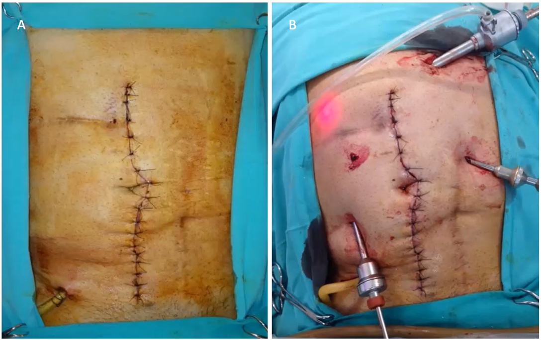
The approach for reoperation in patients with AL is not determined solely by the patient’s clinical condition. Laparoscopic reoperation has a learning curve, which was analysed in a series from Hospital Alemán in Buenos Aires.5 The study analyzed 132 patients who underwent laparoscopic reoperation after laparoscopic colorectal surgery. As the surgical team gained experience, both the conversion rate and operative time decreased. The authors concluded that adequate experience in this setting was achieved after 50 laparoscopic reoperations.
Surgeons must also consider the experience of the assisting surgeon and the availability of laparoscopic equipment. An assistant with limited laparoscopic experience may prolong the procedure and potentially contribute to complications. Many centers in Argentina lack 24–hour access to laparoscopic equipment and video systems. These logistical constraints must therefore be addressed to make laparoscopic reoperation for AL feasible, particularly when emergency surgery is required overnight.
In general, a laparoscopic approach is preferred because of the established benefits of minimally invasive surgery. Postoperative recovery is improved, with less pain and ileus, resulting in a shorter hospital stay.6,7 Surgical site infection, a frequent complication when operating in a septic environment, is less common after laparoscopic reoperation because of reduced wound contamination and smaller incisions. In the long term, this translates into a lower incidence of incisional hernia after laparoscopic reoperation.8
Initially, the use of laparoscopy in patients with peritonitis was controversial because of concerns about the potential spread of infection. Bacterial translocation related to pneumoperitoneum and increased intra–abdominal pressure was thought to potentially exacerbate the systemic inflammatory response.9 However, with increasing surgical experience and technological advances, laparoscopic reoperation has been shown to be feasible and safe in selected patients, allowing adequate identification of the septic source, effective peritoneal lavage, and, in some cases, definitive treatment with less surgical trauma.

Another consideration is the impact that the decision to perform an open reoperation may have on the patient and the potential medicolegal implications. Conceptually, reusing the incision from the initial operation might be associated with a lower perception of additional surgical trauma. Nevertheless, the relationship between the surgical approach and potential medicolegal disputes is multifactorial and depends primarily on the quality of the information provided to the patient, appropriate clinical documentation, and physician–patient communication.
Visceral injuries may occur during laparoscopic reoperation, increasing morbidity and mortality because of adhesions and bowel distention, a common manifestation of AL. Early reoperation clearly makes laparoscopy safer and reduces the risk of complications. Conversion rates for laparoscopic reoperation in patients with AL may exceed 30%, most commonly because bowel distention, which is frequent in this clinical setting, prevents complete exploration of the abdominal cavity.10
One of the earliest published experiences in this field was reported in 2009 by our Colorectal Surgery team at Hospital Alemán in Buenos Aires.11 The study compared outcomes in 27 patients, including 10 who underwent open reoperation and 10 who underwent laparoscopic reoperation. Although the sample size was small, only one previous series had been published on this topic.12 AL was the most common indication for laparoscopic reoperation, and the study demonstrated that a laparoscopic approach was feasible for managing complications after laparoscopic colorectal surgery.
In 2021, the same group published an updated analysis of a substantially larger cohort.13 The study included 159 patients: 124 (78%) underwent laparoscopic reoperation, and 35 (22%) underwent open reoperation. The groups did not differ in demographic characteristics. AL was the most common finding and was successfully managed using a minimally invasive approach. The most frequent strategy consisted of peritoneal lavage, placement of an intra–abdominal drain, and creation of a proximal diverting ostomy. Patients who underwent laparoscopic reoperation had a lower rate of severe complications, whereas mortality was similar between the groups.
Subsequent systematic reviews14,15 have reported consistent findings, further supporting the feasibility and safety of laparoscopic reoperation in patients with AL after colorectal surgery. Compared with open reoperation, the laparoscopic approach is associated with better postoperative recovery outcomes.
The following section describes the surgical technique for laparoscopic reoperation in patients who develop complications during the postoperative period after laparoscopic colorectal surgery.
Patient Positioning and Operating Room Setup
The patient must be securely positioned on the operating table to allow the changes in table position required for systematic exploration of the abdominal cavity. Ideally, both arms should be tucked at the patient’s sides, and a urinary catheter should be placed. Nasogastric or orogastric decompression is often necessary, as gastric decompression facilitates the procedure by improving the operative field.
For left–sided anastomoses, the use of leg supports is important because the Lloyd–Davies position permits perineal access, which is often required to irrigate the rectal stump. A mobile laparoscopic monitor, or one that can be easily repositioned, is recommended so that it can be moved according to the abdominal quadrant being explored. This arrangement provides adequate visualization during lavage of all 4 quadrants, improves operating room ergonomics, and prevents awkward working positions. Proper alignment of the surgeon, operative field, and monitor is essential for efficient exploration, particularly during reoperation for intra–abdominal sepsis, when thorough access to and lavage of the entire abdomen are required. Efficient suction–irrigation systems that allow continuous, targeted lavage are also essential, as are appropriate instruments for careful adhesiolysis.
Abdominal Access and Trocar Placement
Abdominal entry must be performed cautiously. Initially, the sutures closing the midline trocar incision should be removed. Digital dissection can then be performed by introducing a finger through the trocar site and carefully releasing adhesions to allow placement of the first trocar. Once the first trocar is in place, the laparoscope is introduced. Small–bowel distention, which is common in this setting, may make abdominal exploration somewhat more difficult. Changes in operating table position are important for optimizing the operative field and facilitating placement of the remaining trocars. All subsequent trocar insertions must be performed under direct vision to minimize the risk of iatrogenic bowel injury.
Creating a pneumoperitoneum through preexisting abdominal drains has been proposed as an alternative access technique for reoperation in patients with AL, particularly those at high risk of adhesions and distorted anatomy. This approach may allow controlled initial insufflation of the peritoneal cavity, potentially reducing the risk of visceral injury during insertion of the first trocar in a hostile abdomen. However, its applicability is limited because successful pneumoperitoneum depends on drain patency, location, and correct intraperitoneal positioning. Inadequate insufflation, uneven gas distribution, and leakage that compromises effective intra–abdominal pressure may occur. Although this technique may be considered in selected cases by experienced surgeons, it does not replace conventional access methods under direct vision, such as the open technique, which remain the recommended standard for laparoscopic reoperation.
Abdominal Exploration and Peritoneal Lavage
After entering the abdominal cavity, a sample of peritoneal fluid should be obtained for culture and antimicrobial susceptibility testing. Thorough lavage with copious amounts of warm saline is essential. A systematic exploration of the abdominal cavity should be performed to confirm the suspected complication identified by preoperative diagnostic studies and to rule out other potential sources of sepsis. Inadequate lavage increases the risk of residual abscesses and subsequent reoperations, thereby increasing patient morbidity.
If an AL is suspected but not readily apparent, an intraoperative ALT may help identify the defect. As noted above, positioning the patient in the Lloyd–Davies position is therefore important.
Abdominal Wall Closure After Open Reoperation
When an open approach is used, primary abdominal closure is feasible in the vast majority of patients, except in specific circumstances discussed in the section entitled “Open Abdomen and Containment.” The incidence of abdominal wall complications in this setting may reach 40%.16 Fascial and wound complications can have substantial short– and long–term consequences; prevention is therefore a key consideration.
Long–term consequences include discomfort, limitations in occupational and physical activities, chronic pain, abdominal wall deformity, and intestinal obstruction that may require emergency reoperation. Several conditions predispose patients to abdominal wall complications, including diabetes, chronic obstructive pulmonary disease, smoking, obesity, immunosuppression, and previous abdominal surgery.
Before closure, the wound should be irrigated with saline because surgical site infections in this setting are an important determinant of subsequent wound dehiscence and evisceration. Every effort should therefore be made to prevent wound infection.
After laparotomy, abdominal wall specialists recommend the small–bites technique using a continuous suture of 2–0 polydioxanone (PDSⓇ), with a suture–length–to–wound–length ratio of 4:1 (4 cm of suture for every 1 cm of wound).17 No differences in abdominal wall complication rates have been reported between continuous and interrupted sutures. However, the shorter closure time achieved with continuous suturing is sufficient to favor this technique. Peritoneal closure is not recommended because it provides no benefit and prolongs operative time.
The use of intraperitoneal mesh in this setting remains controversial. The World Society of Emergency Surgery guidelines16 note that the published evidence is conflicting and that many surgeons remain reluctant to use mesh in patients with peritonitis. Nevertheless, the guidelines suggest mesh placement because it reduces the incidence of incisional hernia. The optimal mesh type and placement site within the abdominal wall remain controversial.
Recommendations for closure of laparoscopic incisions are straightforward: all trocar–site incisions measuring 1 cm or more should be closed. The recommendations are the same for emergency and elective surgery. Trocar–site hernias are uncommon, with a reported incidence of approximately 0.1% to 1%.
Unfortunately, evisceration remains relatively common in this setting.
Treatment Strategies
If either bowel end is nonviable at reoperation, resection is mandatory. In general, exteriorization of the bowel ends is preferred in this setting, although repeat anastomosis may be considered in selected cases, as discussed below. Once bowel ends viability has been confirmed, the preferred approach is thorough peritoneal lavage, placement of a drain adjacent to the anastomosis, and creation of a proximal diverting loop ostomy. For small defects involving less than 25% of the anastomotic circumference or measuring less than 1 cm, management generally consists of drainage adjacent to the anastomosis and proximal diversion, provided there is no evidence of compromised perfusion. This strategy allows preservation of the anastomosis in situ and may substantially reduce the likelihood of a permanent ostomy, thereby improving long–term quality of life.
One factor to consider when creating a proximal ostomy is the presence of fecal contents in the colon. Intraoperative colonic lavage may help reduce the fecal and bacterial burden in the colon proximal to the diverting ostomy, potentially limiting septic complications in the postoperative period. However, the time required to perform adequate colonic lavage and the technical challenges associated with the laparoscopic approach limit its use in this setting.
Defects involving more than 25% of the anastomotic circumference (or measuring more than 1 cm) are generally considered nonviable. Resection of the affected bowel ends followed by reanastomosis is an option, but its appropriateness in the setting of peritonitis is debatable. These patients are more commonly managed with anastomotic takedown and exteriorization of the bowel ends (Fig. 3). Depending on the anatomical location of the anastomosis, both ends may be brought out together, which is ideal when considering subsequent restoration of intestinal continuity. Leaving closed bowel ends within the abdominal cavity should be avoided whenever possible because failure of the closure can lead to complications and may make subsequent reconstructive surgery more challenging. This issue is discussed in the section entitled “Patients with Hemodynamic Instability.”

Although suturing the anastomotic defect has been described, it is not generally used as the primary treatment strategy. Sutures placed to reinforce the site of the AL are often ineffective because the edges of the defect are typically edematous and friable, making it difficult for the sutures to maintain tension. In addition, there is a substantial risk of enlarging the defect because the surrounding tissue tends to tear in this setting. Nevertheless, despite the limited available evidence, reinforcing sutures may be considered when the defect is very small, the anastomosis is adequately perfused, local contamination is minimal, and a proximal diverting ostomy is created. No prospective studies have compared the use of reinforcing sutures with no suturing when AL is identified intraoperatively. Accordingly, this approach is generally reserved for isolated cases.
Anastomotic reconstruction at reoperation for AL may be considered in hemodynamically stable patients with limited intra–abdominal contamination and viable bowel tissue, allowing resection of the affected segment and creation of a new, tension–free anastomosis. This approach should be reserved for highly selected cases involving early AL, likely caused by a technical defect.
In this regard, if the anastomosis failed under the most favorable physiological conditions available to the patient, that is, at the time of the initial resection, there is little logical basis for assuming that reconstruction under the less favorable clinical conditions resulting from AL will offer a greater likelihood of success. In the series reported by El–Ahmar et al.,18 discussed in greater detail below, the highest mortality rate was observed when colonic resection and repeat anastomosis were performed during reoperation for AL (28.6%), compared with other strategies, including oversewing the anastomotic defect, oversewing combined with proximal diversion, and anastomotic takedown.
The published evidence on anastomotic reconstruction in this setting is very limited and consists primarily of small case series involving colonic resections,19 left colectomies or anterior resections.20 In most cases, proximal diversion is recommended to reduce the risk of recurrent AL. When faced with a small anastomotic defect, the surgeon will likely favor proximal diversion alone over anastomotic reconstruction with proximal diversion. This approach requires less operative time and is technically less demanding, because of tissue edema and adhesions to adjacent structures. Accordingly, repeat anastomosis should be reserved for a highly selected group of patients, particularly those undergoing early reoperation for a presumed technical defect and those with minimal local inflammatory changes. Otherwise, reconstruction may expose patients to the risk of recurrent anastomotic leak. The decision to reconstruct the anastomosis should therefore be individualized and based primarily on intraoperative findings, tissue viability, and the degree of intra–abdominal contamination.
A series published in 2023 analyzed outcomes in 123 patients with AL, including 92 after colonic surgery and 31 after rectal surgery, and compared reoperation outcomes according to the surgical strategy used.18 The main limitations were that the choice of surgical technique was at the discretion of the operating surgeon and that the extent of anastomotic dehiscence was not reported. Nevertheless, the study provides relevant findings, summarized in Table 1. Among patients with colonic and upper rectal anastomoses, oversewing the anastomosis combined with creation of a proximal ostomy was associated with the highest rate of anastomotic preservation.
Table 1. Outcomes of reoperation according to surgical strategy
Anastomotic preservation rate | Number of reoperations | Length of hospital stay (days) | Mortality | |
Colonic Resections | ||||
Primary repair | 37.5% | 2.4 | 26 | 1/8 (12.5%) |
Primary repair with diversion | 77.8% | 1.5 | 26 | 1/18 (5.9%) |
Resection and reanastomosis | 57.1% | 1.6 | 24 | 4/14 (28.6%) |
Anastomotic takedown | 0% | 1.3 | 19 | 8/52 (18.2%) |
Rectal Resections | ||||
Primary repair with diversion | 77.8% | 1.5 | 28 | 0 (0%) |
Washout and drainage | 60% | 1.6 | 13 | 1/5 (20%) |
Anastomotic takedown | 0% | 1.1 | 29 | 1/17 (5.9%) |
Considerations according to the site of anastomotic leakage
Right Colectomy
The surgical management of AL following right colectomy has several distinctive features. First, the available evidence is more limited than that for AL following left colectomy or sigmoidectomy. In addition, experience with laparoscopic management of this complication is more limited. The higher mortality associated with generalized peritonitis secondary to ileocolic AL may represent a relative contraindication to the laparoscopic approach. Vignali et al.21 reported higher rates of generalized peritonitis, hemodynamic instability, and mortality among patients with ileocolic AL than among those with colorectal AL. Another factor that may contribute to this increased mortality is the lower sensitivity and specificity of imaging studies for detecting AL after right colectomy. As discussed in the “Imaging Studies” section, several meta–analyses have confirmed these findings. For example, in 2014, the Dutch Surgical Colorectal Audit group analyzed data from more than 15,000 patients and reported mortality rates following AL of 20.4% after right colectomy and 11.6% after left colectomy.22
For small ileocolic anastomotic defects, the surgeon has several options:
– Repeat anastomosis, with or without proximal diversion.
– Reinforcement or repair of the anastomosis, with or without proximal diversion.
– Proximal diversion without intervention at the anastomotic site.
– Resection of the anastomosis, with the 2 bowel ends brought out together as a double-barreled ileocolostomy or as separate stomas.
Unfortunately, the literature reports substantial variation in the choice of surgical technique. Different leading surgical groups favor different approaches, without a clear rationale for these preferences. For example, Vignali et al.21 analyzed 26 patients with AL among 428 patients who underwent right colectomy. In 61.6% of patients, a small defect (<1 cm) with moderate peritoneal contamination was managed by reinforcing or repairing the anastomosis after lavage of the abdominal cavity with 5 L of warm saline. In the presence of a larger anastomotic defect, the anastomosis was resected and reconstructed with a side–to–side isoperistaltic anastomosis. The decision to create an additional ileostomy was left to the discretion of the operating surgeon. Although the authors did not report the factors guiding this decision, an ileostomy was created in only 22.4% of patients undergoing reoperation. The authors emphasized that early intervention may allow avoidance of an ostomy in most patients, given that AL is likely attributable to a technical defect in the stapled anastomosis, provided there is no evidence of compromised anastomotic perfusion.
In contrast, Dauser et al.3 reported that AL after right colectomy was managed with anastomotic repair and drainage in 15.7% of patients, repeat anastomosis in 37.2%, and ileostomy creation in 47.1%. The surgical strategy was not associated with differences in intraoperative findings, as both MPI and the interval to reoperation were similar across groups. Notably, the likelihood of anastomotic repair at reoperation differed according to whether the initial operation and reoperation were performed by the same surgeon. Anastomotic repair was more frequent when both procedures were performed by the same surgeon (26.5% vs 10.3%). Thus, intraoperative findings did not appear to be the primary determinant of the surgical strategy selected at reoperation.
When an ostomy is created, the surgeon should be aware that restoration of intestinal continuity differs according to the type of ostomy. The rate of AL following ileostomy closure is low, generally less than 2% to 3%, with overall morbidity ranging from 20% to 30%.23 Stapled anastomoses may be associated with a lower rate of postoperative bowel obstruction and shorter operative time, without significant differences in AL rates. In contrast, restoration of intestinal continuity through an ileocolic anastomosis, as occurs during closure of an ileocolostomy, is technically more complex and is associated with a higher complication rate. The rate of AL may reach 8.8%, with an overall morbidity of 27.3%.24 Systematic reviews have reported leak rates of 0% to 8.5% for ileocolic anastomoses, significantly higher than those observed for small–bowel anastomoses. These differences may be explained by the heterogeneous nature of ileocolic anastomoses, including differences in bowel diameter, a greater bacterial burden, and variations in vascular supply. In the absence of direct comparative studies, the available evidence suggests a higher risk of septic complications with ileocolic anastomoses.
Transverse Colectomy/Splenic Flexure Segmental Resection
As discussed in the preceding sections, for small anastomotic defects, the surgical team must decide whether to leave the anastomosis in situ and perform a diverting ostomy or resect the anastomosis and bring out both bowel ends as stomas. The choice between these strategies depends largely on the size of the defect, as described above.
However, it is important to note that restoration of intestinal continuity after loop colostomy reversal is technically simpler, typically requiring dissection limited to the ostomy site, and is associated with low morbidity. In contrast, reversal of an end colostomy requires a formal colonic anastomosis in the setting of a defunctionalized distal colon, entailing greater technical complexity, a higher risk of septic complications, and a longer functional recovery. Additionally, the defunctionalized distal limb or mucous fistula generally undergoes atrophy and becomes smaller than the proximal limb. This disparity in bowel caliber may complicate end–to–end anastomosis and require a side–to–side configuration.
Sigmoidectomy/Low Anterior Resection
When a colorectal anastomosis cannot be salvaged, a temporary end colostomy should be created in the left lower quadrant, with closure of the rectal stump, as the rectal remnant is too short to be brought out through the abdominal wall. Thorough irrigation of the rectal stump is essential because, as previously discussed, it reduces the bacterial burden and fecal contamination, thereby mitigating the septic consequences of rectal stump closure failure. In addition, nonabsorbable marking sutures should be placed to facilitate identification of the rectal stump during subsequent reconstructive surgery.
A key consideration with this approach is the high rate of permanent stoma formation. For several reasons, Hartmann reversal is never performed in approximately 50% of patients. Therefore, whenever feasible, preserving the anastomosis and creating a proximal diverting ostomy is preferable. This strategy substantially increases the likelihood of restoring intestinal continuity and reduces the rate of permanent stoma formation to approximately 15%.3
Available evidence from randomized controlled trials and meta–analyses has not demonstrated a clear superiority of loop ileostomy over loop colostomy, or vice versa, for the management of ALs. However, loop ileostomy is consistently associated with fewer complications, particularly stoma prolapse, which has led to its preferential use in contemporary practice.25
When the anastomosis has been preserved with a proximal diverting ostomy, anastomotic stricture must be ruled out before stoma reversal. A contrast study can identify a contained AL or, alternatively, an anastomotic stricture. AL triggers a local inflammatory response characterized by the release of cytokines and growth factors. When this process is prolonged or exacerbated, persistent activation of fibroblasts and myofibroblasts increases collagen synthesis and deposition, leading to circumferential fibrosis and progressive loss of bowel wall elasticity. The end result is anastomotic stricture.
Endoscopic dilation should be performed first to ensure an adequate luminal diameter at the anastomotic site. If malignancy is suspected, biopsies should be obtained. This issue is discussed further in the section entitled “Chronic AL.”
Low Anterior Resection with a Diverting Ostomy
As discussed in the preceding section, in the setting of extraperitoneal rectal anastomoses, every effort should be made to preserve the anastomosis in situ. When the anastomosis is taken down and the rectal stump is closed, restoration of intestinal continuity is achieved in only a small proportion of patients, substantially increasing the likelihood of a permanent stoma. The likelihood of successful restoration of intestinal continuity is directly proportional to the length of the remaining rectal stump.
In certain circumstances, compromised anastomotic viability necessitates resection. Depending on the patient's overall condition, reanastomosis may be considered at this point. However, this may be technically challenging because of the limited length of the remaining rectal stump. In selected patients, a delayed coloanal pull–through procedure represents a viable alternative and is discussed in detail in the section entitled “Chronic AL.” An important limitation, however, is that this approach requires a lengthy and technically complex operation, as transperineal exteriorization of the colonic limb necessitates extensive colonic mobilization and, in most cases, splenic flexure mobilization.
Ileal Pouch–Anal Anastomosis (IPAA)
The two most common indications for IPAA are ulcerative colitis and familial adenomatous polyposis (FAP). The reported incidence of AL after IPAA ranges from 5% to 15%, with rates varying according to the underlying indication for surgery. Among patients with ulcerative colitis, the leak rate exceeds 20% in some series, reaching 23.4%, whereas in patients with FAP, it remains below 10%.26 These differences are likely attributable to the poorer overall condition of patients with ulcerative colitis, including malnutrition and immunosuppressive therapy, which are common in this population.
The clinical consequences of AL after IPAA range from severe sepsis to mild, chronic manifestations. Management depends on the timing of presentation and the severity of symptoms.27
In patients with uncontrolled sepsis, the initial approach should follow the principles outlined in the section “The Hemodynamically Unstable Patient.” If a diverting loop ileostomy is not already in place, proximal diversion should be the first step. This is particularly relevant because some centers advocate IPAA without a protective ostomy. In patients with persistent fecal contamination despite proximal diversion, closure of the efferent limb should be considered. Pelvic sepsis requires aggressive management, with drainage via percutaneous, transanal, or transabdominal approaches, the latter when surgical reexploration is necessary. Adequate drainage is essential for resolution of pelvic sepsis, and patients should be counseled that multiple interventions may be required and that treatment may extend over several months. Transanastomotic drains may be placed and progressively exchanged for smaller–caliber drains during subsequent procedures. For presacral cavities, several previously described options are available, including negative–pressure therapy and cavity opening using stapling devices, energy–based instruments, or endoscopic techniques.
In some cases, AL may manifest as a pouch–vaginal fistula. Although this complication is generally attributable to a technical defect during IPAA construction, Crohn's disease should be ruled out as an underlying cause. Treatment success rates are low, at less than 50%, and options include a mucosal advancement flap or pouch reconstruction.
Dehiscence of the staple line at the tip of the J–pouch is another potential complication. It frequently goes undetected on contrast studies performed before ileostomy closure. Percutaneous drainage is the initial treatment of choice; if symptoms persist, repeat ileostomy may be necessary. When the defect is identified endoscopically, OTSC placement may be a reasonable alternative, with acceptable success rates. Nevertheless, many patients require surgical reexploration and resection of the pouch tip using a stapling device. The staple line should be oriented at a 45–degree angle, with the tip positioned on the mesenteric side, to prevent ischemia that may have contributed to the complication.
When the previously described approaches fail, options include pouch reconstruction or pouch excision with creation of a permanent end ileostomy.
AL after IPAA can substantially compromise pouch function, with direct consequences for continence and quality of life.
In a 2025 multicenter European study by the Dutch MIRACLE group, involving 411 patients from six centers, outcomes after AL following IPAA were analyzed according to whether treatment was initiated early (within 21 days of pouch construction) or late. With early treatment, 90% of pouches were preserved, and 95% of patients were stoma–free at one year after surgery. In contrast, with delayed treatment, pouch preservation was achieved in 60% of patients, and only 40% were stoma–free.28 These differences may reflect the effects of prolonged local sepsis, which can lead to fibrosis and increase the risk of pouch failure.
Although this is an important clinical issue, a detailed discussion of the management of AL after IPAA and its functional consequences is beyond the scope of this review.
Ostomies
Whenever possible, potential ostomy sites should be marked on the abdominal wall preoperatively. Ideally, this should be performed by an enterostomal therapy nurse before reoperation, provided the patient's clinical condition allows.
In some cases, the ostomy may be created through an incision from the previous operation, such as the incision used to extract the specimen. However, this approach should be avoided when the incision is substantially larger than the intended ostomy, as this may lead to postoperative complications. Ostomy appliance management can be challenging in these circumstances, and the wound may break down because of the moist environment created by the ostomy appliance and fecal contamination. Local infections can result in significant morbidity and may occasionally necessitate reoperation to relocate the ostomy. Accordingly, the decision should be individualized by the operating team based on multiple factors, as creation of the ostomy through a preexisting incision is not always feasible.
Once proximal diversion has been deemed necessary, the surgeon must choose between an ileostomy and a colostomy.
Although no studies have specifically compared ileostomy versus colostomy exclusively in the setting of reoperation for AL, the available evidence suggests that loop ileostomy offers advantages as a diversion strategy.
Loop ileostomy is currently the preferred approach in most reoperations for AL, with colostomy reserved for selected circumstances. Stoma prolapse is more common with colostomies for several reasons:29
– The mesocolon is longer and more lax than the terminal ileal mesentery.
– The more solid colonic contents generate more forceful peristaltic waves.
– The larger diameter of the colon requires a larger fascial opening.
Conversely, ileostomy management can be more challenging because of the increased risk of dehydration, particularly in older patients. Ileostomies also leave a greater intraluminal fecal content between the stoma and the site of the AL. In patients too frail to tolerate an ileostomy or whose colon contains a substantial fecal burden, the surgeon may opt for proximal colostomy. Mobilization of the transverse colon may pose technical challenges in some cases; nevertheless, a proximal transverse colostomy remains a suitable option in selected patients.
A distal transverse colostomy, however, may complicate subsequent restoration of intestinal continuity. The blood supply to the mobilized colon may be jeopardized if the inferior mesenteric artery was ligated high during the index operation or if the marginal artery supplying that segment was inadvertently divided. Accordingly, in patients with colorectal AL, a proximal diversion upstream of the middle colic artery is a reasonable preference.
Loop ileostomy and loop colostomy reversal also differ in several important respects. In general, loop ileostomy closure is technically less demanding because the ileum typically has a smaller caliber, a lower bacterial burden, and less local inflammation, facilitating bowel handling and anastomosis construction. Dissection of the ileal loop is also generally easier because adhesions tend to be less extensive. In contrast, loop colostomy closure can be more challenging because of the larger colonic diameter, greater bacterial colonization, and tendency toward denser adhesions, increasing the risk of intraoperative contamination and infectious complications. Greater tension at the colonic anastomosis and variability in its blood supply may also contribute to complications such as anastomotic leakage and wound infection. Overall, these differences make loop ileostomy closure a technically simpler procedure, generally associated with lower morbidity than loop colostomy closure.
The greater bacterial burden associated with colostomies also contributes to a higher incidence of surgical site infection, which is directly correlated with the risk of incisional hernia at the stoma closure site.29
Hemodynamically Unstable Patient
In the setting of AL, hemodynamic instability is defined as the inability of the cardiovascular system to maintain adequate tissue perfusion as a result of intra–abdominal sepsis.30 From a pathophysiologic standpoint, AL may trigger a systemic inflammatory response secondary to peritoneal contamination and bacterial translocation, leading to vasoplegia, increased capillary permeability, decreased venous return, and sepsis–induced myocardial dysfunction. If timely source control is not achieved, this process may progress to septic shock.
In patients with AL, hemodynamic instability is clinically defined by the presence of 1 or more of the following criteria:31
– Systolic blood pressure <90 mmHg or mean arterial pressure <65 mmHg.
– Requirement for vasopressors to maintain adequate perfusion pressure despite fluid resuscitation.
– Elevated serum lactate level (>2 mmol/L) as a marker of tissue hypoperfusion.
– Oliguria (<0.5 mL/kg/h).
– Altered mental status attributable to hypoperfusion.
– Peripheral signs of poor perfusion, including delayed capillary refill, cool skin, or mottling.
In this setting, hemodynamic instability is a major indicator of disease severity and a key determinant of surgical decision–making. Its presence favors a damage–control approach. From a prognostic standpoint, hemodynamic instability at the time of AL diagnosis is associated with higher mortality, increased reintervention rates, and a lower likelihood of subsequent restoration of intestinal continuity.32
The primary goal of surgery is to achieve adequate sepsis control and stabilize the patient. In some cases, transfer to the ICU may be necessary to initiate appropriate resuscitation with intravenous fluids and antibiotics, together with close monitoring of clinical parameters. Initial management focuses on restoring tissue perfusion through intravenous fluid resuscitation, preferably with balanced crystalloids, guided by clinical findings and dynamic measures of fluid responsiveness.30,31 In patients with persistent hypotension or signs of hypoperfusion despite adequate volume resuscitation, vasopressors may be initiated early, with norepinephrine as the first–line agent to achieve an adequate mean arterial pressure. Broad–spectrum intravenous antibiotics should be administered promptly and subsequently tailored according to culture results. Microbiologic specimens should be obtained without delaying antimicrobial therapy. Monitoring should include serum lactate, urine output, and hemodynamic parameters to guide resuscitation and assess the response to treatment. Nevertheless, the cornerstone of management is control of the septic source, which in this setting requires early surgical reintervention. Hemodynamic resuscitation, antimicrobial therapy, and source control should therefore be considered simultaneous and interdependent interventions aimed at improving patient outcomes.30
If hemodynamic stabilization is achieved, imaging, preferably CT, may be obtained. If hemodynamic instability persists, the patient should be taken to the operating room without delay.
Hemodynamic instability is an absolute contraindication to a laparoscopic approach. In this setting, a midline laparotomy should be performed, with the incision extended according to the clinical suspicion and intraoperative findings. A wide, preferably xiphopubic, laparotomy is often necessary to allow complete exploration of the abdominal cavity. This approach facilitates adequate peritoneal lavage. Broad exposure also helps reduce operative time and optimize ostomy construction, because appropriate stoma placement often requires sufficient mobilization of the corresponding bowel segment.32
Particular care must be taken during entry into the abdominal cavity to avoid inadvertent bowel injury, a complication reported in 5% to 20% of patients in this setting and associated with adhesions, bowel wall edema, and distorted anatomy. If an injury is not recognized during reoperation, the consequences may be severe, including enteric fistulas, interloop abscesses, and increased morbidity and mortality.31
The risk of inadvertent small–bowel injury during relaparotomy is closely related to the timing of reoperation. In general, during the first 3 to 5 days, adhesions are loose and relatively easy to dissect, reducing the risk of iatrogenic injury. By contrast, during later reinterventions, particularly after postoperative days 5 to 7, adhesions become denser and more fibrotic, distorting the anatomy and making tissue planes more difficult to identify. This significantly increases the risk of bowel injury during abdominal entry or adhesiolysis.33 Persistent sepsis and multiple prior laparotomies further complicate reoperative surgery by increasing the technical difficulty of abdominal re–exploration.
Accordingly, when reintervention is indicated, an early approach not only facilitates source control but may also reduce the risk of inadvertent bowel injury and its associated complications.
Once the abdomen has been entered, the first step should be a thorough exploration of the abdominal cavity to identify the source of sepsis. After the source has been identified, a sample of intra–abdominal fluid should be obtained for culture, followed by peritoneal lavage. Lavage should be performed systematically and thoroughly using warm saline, with careful inspection and irrigation of all abdominal compartments, including the subphrenic spaces, paracolic gutters, and pelvis. Adequate mobilization of the bowel loops is essential to avoid residual interloop abscesses. Intraperitoneal antiseptic or antibiotic solutions have not demonstrated consistent benefits and are not recommended for routine use.
In general, the anastomosis should be resected in this setting, as the defect is likely to be extensive, and both bowel ends should be brought out as stomas.32 Whenever feasible, both ends should be brought out adjacent to one another, as this facilitates subsequent reconstruction. However, if mobilization of the bowel ends would substantially prolong the operation, the stomas may be matured at separate abdominal sites. As a last resort, in selected patients whose overall condition does not permit the additional operative time required for dissection and stoma construction, the nonfunctioning distal bowel end may be closed and left within the abdominal cavity. This strategy is not without complications and should therefore be reserved for carefully selected patients. Ischemia of the closed bowel stump is not uncommon and may result in closure failure and subsequent peritonitis. Furthermore, subsequent operations are likely to make adequate bowel mobilization for mucous fistula creation increasingly difficult. Failure to mobilize the bowel during the initial operation may therefore compromise future reconstructive options.32
For ileocolic and colocolic anastomoses, the anastomosis should be resected and, whenever feasible, both bowel ends should be brought out to the abdominal wall, as described above. If the dissection required to accomplish this would prolong the operation, a proximal ostomy may be created and a drain placed at the anastomotic site.
For colorectal anastomoses, the anastomosis should also be resected, with creation of an end colostomy and closure of the rectal stump. Ideally, the rectal stump should be closed with a stapling device, as hand–sewn closure may substantially prolong the operation. Intraoperative rectal stump lavage is important because it may prevent serious complications. It generally adds little operative time and should be avoided only in patients with severe hemodynamic instability. Rectal stump lavage may mitigate the consequences of closure failure by reducing the bacterial burden and fecal contamination, thereby limiting the septic impact of a subsequent leak. Conversely, if the stump dehisces in the presence of a substantial fecal load, the resulting contamination may lead to pelvic peritonitis and subsequent generalized peritonitis.33
In some cases, dissection at the site of AL may be extremely challenging because of adhesions to adjacent structures. Dissection in this area may result in complications even more severe than those associated with AL itself. In such circumstances, thorough peritoneal lavage, placement of a drain at the site of AL, and creation of a proximal ostomy are mandatory.
Once the acute process has been addressed, the operating team must decide whether to close the abdomen or defer definitive closure, leaving the abdomen open with temporary abdominal containment. The latter approach is discussed in the following section.
Open Abdomen Management with Temporary Abdominal Closure (TAC)
In certain circumstances, particularly in patients with extensive peritonitis and severe physiologic derangement, multiple reoperations may be required to achieve adequate sepsis control. In this setting, leaving the abdomen open with TAC may appear to be a reasonable strategy to facilitate planned relaparotomies every 24 to 48 hours. Although this approach can be lifesaving in critically ill patients, it is associated with serious complications, and approximately 25% of patients managed with an open abdomen will develop an enteric fistula.34,35 The role of this strategy remains controversial, as adequate peritoneal lavage may obviate the need for an open abdomen.
The concept of the open abdomen was initially adopted as a damage–control strategy in military surgery. Experience in this setting showed that patients could have better outcomes when the initial operation was abbreviated to achieve hemorrhage control or initial sepsis control, leaving the abdomen open for subsequent definitive surgery once hemodynamic stability had been restored. These principles were subsequently extrapolated to the management of postoperative intra–abdominal sepsis, in which AL may be a frequent cause.
The role of open abdomen management in intra–abdominal sepsis has evolved considerably over the past several decades. Initially used extensively to facilitate source control and allow planned re–explorations, its indications have progressively narrowed as the associated complications have become better recognized. These include loss of abdominal domain, enteroatmospheric fistulas, persistent infection, and difficulties achieving definitive abdominal closure. Current consensus increasingly supports the use of the open abdomen as a selective intervention reserved for specific clinical circumstances rather than as a routine management strategy (Fig. 4).36
Effective source control combined with adequate resuscitation has made primary abdominal closure feasible in many patients, even in complex septic scenarios, thereby reducing the need for an open abdomen. Current indications are therefore driven primarily by technical or physiologic constraints rather than by sepsis alone. These include elevated intra–abdominal pressure, significant visceral edema, inability to achieve tension–free fascial closure, and the risk of or established abdominal compartment syndrome.36 In these circumstances, the open abdomen serves primarily as a decompressive strategy to preserve visceral perfusion rather than as a means of achieving septic source control. Although temporary abdominal closure facilitates reintervention and ongoing management of the septic source, indiscriminate use may perpetuate inflammation, promote fluid and protein losses, and increase morbidity and mortality. Accordingly, current practice favors an individualized approach that prioritizes early abdominal closure whenever feasible and reserves the open abdomen for patients in whom safe closure cannot be achieved. This shift reflects an improved understanding of the pathophysiology of intra–abdominal sepsis and the detrimental effects of an open abdomen, supporting a more balanced approach that integrates source control, physiologic stability, and preservation of abdominal wall integrity. Therefore, whenever the patient's clinical condition permits, the abdomen should be closed following emergency laparotomy in this setting.
In some patients with AL, tissue edema associated with the frequently encountered ileus may increase intra–abdominal pressure, making closure under tension potentially hazardous. In these circumstances, temporary abdominal closure is preferred to avoid the consequences of increased intra–abdominal pressure, impaired visceral perfusion, renal dysfunction, increased intrathoracic pressure, compromised mechanical ventilation, and progression to abdominal compartment syndrome.
The diagnosis of intra–abdominal hypertension relies on objective measurement of intra–abdominal pressure, with intravesical pressure measurement being the most widely used method in clinical practice. According to the World Society of the Abdominal Compartment Syndrome, intra–abdominal hypertension is defined as a sustained or repeated intra–abdominal pressure of ≥12 mmHg.37
Several commercial devices are available for temporary abdominal closure. However, in Argentina, locally assembled, lower–cost systems are preferred because of financial constraints. The most commonly used system consists of a urinary drainage bag that is opened and fenestrated, a sponge, a suction tube, which may be a nasogastric tube, and a waterproof adhesive drape, such as Steri–Drape®. The system is then connected to suction, allowing effective compression of the dressing. Initial concerns regarding an increased incidence of enteroatmospheric fistulas have been raised; however, negative pressure levels below 125 mmHg have not been associated with this complication.38
In some cases, repeat abdominal washouts every 48 to 72 hours are required to achieve adequate source control. However, fascial closure may become technically challenging after 72 hours. Without appropriate measures to maintain fascial approximation, definitive abdominal closure may become virtually impossible by postoperative day 10. In this regard, the 2018 European Hernia Society guidelines favor early dynamic closure over static closure.17 Dynamic closure involves placing a nonabsorbable mesh secured with a running suture to the fascial edges of the abdominal wall. At each subsequent return to the operating room, the mesh is opened along the midline to allow surgical re–exploration and is then reapproximated with a running suture, progressively bringing the fascial edges toward the midline. With each successive washout, gradual tension is applied during closure, facilitating progressive medialization of the fascial edges. Definitive fascial closure is generally achieved by the third or fourth reoperation, at which point the mesh can be removed and the fascia closed (Figs. 5 and 6).
CHRONIC ANASTOMOTIC LEAKAGE
Chronic AL is defined as leakage that becomes apparent more than 30 days after surgery. The literature primarily addresses AL after LAR and IPAA, as both procedures commonly involve a diverting stoma that may delay the clinical presentation of AL in the early postoperative period. In this setting, approximately 50% of leaks that develop during the first few postoperative days become chronic within the first postoperative year, underscoring the importance of appropriate management.39
Chronic AL may present in 2 distinct settings: persistent leakage from the original anastomosis or leakage developing after closure of a diverting stoma. Clinical manifestations vary considerably, ranging from minimally symptomatic disease to exceptionally severe complications, including osteomyelitis, necrotizing fasciitis, complex fistulas, hydronephrosis secondary to ureteral compression, chronic pain, and persistent fever.

In patients who have undergone surgery for malignancy, associated local recurrence must be ruled out through targeted diagnostic evaluation and, when indicated, histopathologic examination.
Most cases of chronic AL result from persistence of an acute leak recognized during the early postoperative period. In some patients, however, diagnosis is delayed, and presentation may be nonspecific, with symptoms such as malaise, anorexia, fever, or purulent drainage from the perianal region or anal canal.39
The initial evaluation should include a proctologic examination. Inspection is performed to identify fistulas and collections. Digital rectal examination may be painful and uncomfortable in this setting, but when feasible, it can reveal an anastomotic defect. Anoscopy allows direct visualization of the anastomosis. However, given the pain frequently associated with proctologic examination in these patients, examination under anesthesia is an important alternative. A small posterior anastomotic opening is commonly identified, through which purulent fluid may drain.
A contrast study performed by instilling contrast through the anus is useful for delineating the fistulous tract and the underlying cavity. Particular care must be taken when performing this study, because catheter placement proximal to the defect or inflation of the catheter balloon at the anastomotic level may compromise the examination. Additional imaging modalities include CT and pelvic MRI, which help define the relationship between the cavity or fistula and the sacrum and adjacent organs.
Several therapeutic options are available and may be used in combination in selected cases.39
1) Fibrin Glue
The use of fibrin glue for this indication was first described in 2004. The fistulous tract is initially curetted, followed by instillation of fibrin glue.40 Although Swain et al reported a 100% success rate in their series, this result was not reproduced in subsequent studies, and the clinical utility of this approach remains controversial.
2) OTSC
The OTSC technique was described in previous sections. Case series have reported a success rate of approximately 80% in this setting, although evidence from higher–quality studies remains limited.
3) Marsupialization of the Presacral Cavity
The goal of this procedure is to establish communication between the presacral cavity and the rectal lumen. Several techniques have been described, including the use of laparoscopic stapling devices and energy–based instruments. Once the cavity has been opened into the rectal lumen, debridement of the intracavitary tissue is recommended whenever feasible to promote adequate healing. After the cavity has healed, intestinal continuity can be restored. Subsequent defecatory dysfunction may occur. The available literature on this technique remains limited to case series.
4) Minimally Invasive Techniques
Transanal endoscopic microsurgery (TEM) and TAMIS have been reported as useful approaches in recent years. Both techniques provide direct visualization of the anastomotic defect through a transanal approach.
The rectum is circumferentially divided through its full thickness, and the proximal bowel is closed above the transection site to exclude it from the dissection plane. Simultaneously, transabdominal dissection is performed toward the pelvis using either an open or minimally invasive approach. Transanal dissection proceeds proximally until it meets the abdominal dissection, allowing mobilization of the failed anastomosis. Once the 2 dissection planes meet and the colon has been adequately mobilized, the colon is brought down to create a new anastomosis, followed by construction of a proximal diverting stoma.
TAMIS has been reported to achieve restoration of intestinal continuity in 76% of patients.41
5) Anastomotic Resection
In selected patients, more extensive surgery may be required to restore intestinal continuity. Preoperative anorectal manometry can help assess sphincter function, as patients with significant sphincter dysfunction are poor candidates for reconstructive procedures of this type.
In these procedures, dissection within a hostile pelvis characterized by inflammatory adhesions and fibrosis can be particularly challenging. Ureteral catheter placement may be helpful to facilitate ureteral identification and prevent injury or enable its intraoperative recognition.
An open approach appears to be the most appropriate option given the technical challenges of reoperative pelvic surgery in the presence of inflammatory adhesions and fibrosis.
One reconstructive option is a Soave–type coloanal pull–through, in which the colon is brought down through the muscular cuff of the rectal stump (endorectal pull–through), followed by creation of a coloanal anastomosis.
When immediate reconstruction is not feasible, usually because of poor tissue quality, delayed coloanal anastomosis using a pull–through technique is a valid alternative. This technique was described for the treatment of megacolon in 1961 by 2 surgeons working independently: Robert Turnbull of the Cleveland Clinic,42 who used it in patients with rectal cancer and Hirschsprung disease, and Daher Cutait of São Paulo,43 who used it in patients with Chagas–related megacolon. The procedure is therefore known as the Turnbull–Cutait pull–through technique.
Although its use declined with the development of stapling devices, numerous case series published over the past 20 years have described its utility in managing a hostile pelvis resulting from inflammatory adhesions and fibrosis associated with AL. The procedure has been used in a variety of settings, including benign and malignant disease, radiation–related complications, rectovaginal and rectoureteral fistulas, complex fistulas associated with Crohn disease, and selected neoplastic conditions.44
In patients with AL, the procedure is generally performed in 3 stages because most already have a diverting stoma. The stages are as follows: 1) colonic mobilization and transanal pull–through, leaving a segment of colon exteriorized; 2) resection of the exteriorized segment and delayed hand–sewn coloanal anastomosis approximately 7 to 10 days later; and 3) closure of the diverting stoma.
One of the main advantages of this procedure in elective surgery is the potential to avoid a diverting stoma. However, it may seem paradoxical that most published series involving emergency surgery favor combining the technique with a proximal diverting stoma. This difference likely reflects the distinct clinical circumstances of emergency surgery, in which surgeons tend to adopt a more conservative approach. An ileostomy is generally preferred because a colostomy may complicate reoperation for complications of the delayed coloanal pull–through and, in some cases, may preclude the creation of a new anastomosis.
The first stage involves pelvic dissection down to the levator muscles, followed by resection of the previous anastomosis. The colon to be pulled through must be adequately mobilized. Accordingly, complete mobilization of the splenic flexure and ligation of the inferior mesenteric vein at the inferior border of the pancreas are required in the vast majority of cases. In some patients, optimal colonic mobilization requires passage of the colon through a retroileal transmesenteric window, through an avascular plane beneath the ileocolic vascular pedicle. Another option is the Deloyers procedure, which involves mobilization of the hepatic flexure and counterclockwise rotation of the ascending colon.
Through the transanal approach, often with the assistance of a Lone Star® retractor, a mucosectomy of the rectoanal stump is performed, beginning at the dentate line and proceeding proximally. The colonic segment is then delivered through the anus, leaving at least 5 cm of redundant bowel exteriorized. Cardinal sutures of absorbable material are placed to prevent retraction or displacement of the pulled–through colon. The exposed end is covered with petroleum jelly–impregnated gauze. Hospitalization is generally preferred, although selected patients may be discharged with outpatient follow–up.
The second stage is performed approximately 7 days later. The exteriorized segment is resected, and a hand–sewn coloanal anastomosis is created using interrupted absorbable sutures. This interval allows adhesions to develop between the anal margin and the colonic serosa during the first postoperative week. At this stage, it is essential to avoid disrupting any adhesions that have formed between the colonic serosa and the anal canal. Transection should be performed at the level of the anoderm. A more distal transection may result in prolapse, whereas a more proximal transection may require disruption of the adhesions, potentially promoting AL.
Although variations of the procedure have been described, the technique outlined above is the most commonly reported. When a diverting stoma can be omitted, the interval between exteriorization and resection may range from 5 to 14 days.
In patients with a proximal diverting stoma, the third stage consists of stoma closure. A contrast study is obtained before this procedure to rule out an anastomotic defect.
The main causes of failure of this technique are ischemic necrosis, tumor recurrence, and anal stenosis. Ischemia of the pulled–through colon is more common in obese male patients because of vascular compromise associated with delivering the colon through a narrow pelvis. The reported rate of permanent stoma ranges from 1% to 25%. The most common complications are anal stenosis (17%), fistula (7%), colonic prolapse (7%), and AL (3%).45
Published data on resective techniques for chronic AL remain limited. Studies comparing immediate and delayed coloanal anastomosis are difficult to interpret because the patient populations generally differ between groups. Nevertheless, a study published in 2022 by colorectal surgeons at the Cleveland Clinic is noteworthy.46 This single–institution retrospective study compared 81 patients who underwent immediate anastomosis with 129 who underwent delayed anastomosis; most underwent reoperation for rectal cancer and anastomotic complications.
Restoration of intestinal continuity was achieved in 70% of patients in the delayed anastomosis group over a mean follow–up of 1.4 years. The restoration rates at 3 and 5 years were 60% and 50%, respectively. The leading cause of treatment failure was fecal incontinence, which resulted in a persistent stoma or the need for a new stoma.
Fecal incontinence in this patient population is multifactorial, reflecting the interaction of preoperative, technical, and postoperative factors. Preoperative contributors include preexisting sphincter dysfunction, pelvic neuropathy, and reduced rectal compliance, particularly in patients who have received radiation therapy. Technical factors may include injury to the sphincter complex or autonomic innervation during dissection. In addition, the characteristics of the pulled–through colon and its limited adaptation as a neorectum may result in increased stool frequency and urgency, with symptoms consistent with low anterior resection syndrome. Finally, postoperative complications, such as AL and stenosis, may lead to fibrosis and reduced tissue compliance, further compromising function.
Another study comparing delayed and immediate anastomosis is the systematic review and meta–analysis by Galletti et al.47 Postoperative complications were more frequent in the delayed anastomosis group than in the immediate anastomosis group (51% vs 35%), whereas fecal incontinence was less frequent (18% vs 28%). However, at the end of follow–up, the proportions of patients who were stoma–free were similar between groups (81% in the pull–through group vs 83% in the immediate anastomosis group). One of the studies included in the meta–analysis reported a median Wexner fecal incontinence score of 10 (range, 0–20) at 6 months after stoma closure. Overall, more than half of the patients experienced severe or very severe impairment of fecal continence. These findings highlight that restoration of intestinal continuity does not necessarily translate into good functional outcomes or quality of life.
Similar findings were reported by Tan et al.48 in a 2025 systematic review including 97 patients who underwent a delayed Turnbull–Cutait pull–through procedure for AL. The proportion of patients who were stoma–free at 2 years was relatively high, with intestinal continuity restored in 81.4% of patients.
6) Abdominoperineal Resection
In selected patients, severe pelvic fibrosis may preclude the reconstructive procedures described above, particularly when acceptable functional outcomes are unlikely. In these circumstances, abdominoperineal resection with permanent colostomy may be an appropriate option.
Patients in whom a permanent stoma is maintained with the rectum left in situ may continue to experience symptoms such as persistent drainage or chronic pain, in addition to difficulties with adequate endoscopic surveillance. In these circumstances, abdominoperineal resection may also be considered. However, surgery in a heavily fibrotic pelvis can be associated with substantial morbidity because of the technical challenges of dissection. Its use in this setting should therefore be reserved for carefully selected patients.39
APPENDIX. ANTIBIOTIC THERAPY FOR GRADE C ANASTOMOTIC LEAKAGE
Antibiotic therapy should be established promptly, as administration within the first hour of ICU admission has been associated with improved survival.1 In hemodynamically unstable patients, obtaining blood cultures before initiating antimicrobial therapy is essential whenever feasible to help identify the causative pathogens. Unlike the antibiotic strategies discussed in previous sections for patients eligible for nonoperative management, patients with grade C AL generally present with septic shock. Several antimicrobial regimens are available, as outlined below. Empiric antifungal therapy should also be considered in this setting.49
• Meropenem, with or without linezolid, vancomycin, or daptomycin.
• Meropenem, with or without tigecycline.
• Piperacillin–tazobactam, with or without gentamicin or amikacin, and with or without linezolid, vancomycin, or daptomycin.
• Ceftolozane–tazobactam or ceftazidime–avibactam plus metronidazole, with or without linezolid, vancomycin, or daptomycin.
• Ceftolozane–tazobactam or ceftazidime–avibactam plus tigecycline or eravacycline.
• Meropenem–vaborbactam, with or without linezolid, vancomycin, or daptomycin.
Dosing should be individualized according to each patient's clinical condition and relevant clinical parameters. Concomitant Candida spp. infection is common in critically ill patients and is associated with poor outcomes. Accordingly, empiric antifungal therapy is recommended in hemodynamically unstable patients, with echinocandins (anidulafungin, caspofungin, or micafungin) preferred over fluconazole.49 If intra–abdominal fluid cultures are negative for Candida spp., antifungal therapy may be discontinued. If a fluconazole–susceptible Candida species is identified, treatment may be de–escalated to fluconazole. A 10– to 14–day course of antifungal therapy is generally appropriate.50
Intravenous antibiotic therapy for 4 to 5 days is appropriate in hemodynamically stable patients who have achieved adequate source control. In critically ill patients, treatment may be extended to 8 days. When cultures confirm that the causative bacteria are not multidrug resistant, antimicrobial therapy should be de–escalated and tailored to the susceptibility results.51
In this setting, diagnostic imaging is mandatory whenever the patient's hemodynamic status permits.
Although most patients with AL require surgical treatment, the likelihood of reoperation depends on several factors. First, patients with a proximal diverting stoma are more likely to achieve successful resolution of AL with medical and/or percutaneous treatment. Second, the timing of AL onset is an important consideration. The earlier the clinical manifestations develop, the more likely reoperation will be required. Conversely, leaks presenting later in the postoperative course, typically between postoperative days 7 and 10 or thereafter, are more likely to be managed conservatively.52
The reported mean time to reoperation for this complication varies considerably across studies; however, a peak appears to occur on postoperative days 4 and 5.32
CONCEPTUAL SUMMARY
Surgical management of AL should be individualized according to the patient's hemodynamic status, anastomotic location, defect size, and extent of intra–abdominal contamination.
Early reoperation facilitates source control, preserves the feasibility of minimally invasive surgery, and reduces morbidity.
In selected patients, relaparoscopy is a safe alternative associated with faster recovery and lower abdominal wall morbidity.
When the anastomosis is viable and the defect is limited, management should prioritize anastomotic preservation, adequate source control, and proximal fecal diversion. Conversely, extensive defects, ischemia, or hemodynamic instability generally warrant anastomotic resection and creation of an end stoma or stomas. Immediate reconstruction is reserved for highly selected cases. In patients with septic shock or hemodynamic instability, a damage–control approach is warranted, prioritizing expeditious source control, anastomotic resection when indicated, and fecal diversion. In this setting, laparotomy is the preferred surgical approach.
An open abdomen with temporary abdominal closure should be reserved for exceptional circumstances, such as intra–abdominal hypertension, abdominal compartment syndrome, or inability to achieve safe abdominal closure, with early fascial closure prioritized whenever feasible.
Chronic AL requires a stepwise approach that may include endoscopic interventions, drainage procedures, transanal techniques, and reconstructive surgery to restore intestinal continuity and avoid a permanent stoma.
REFERENCES
1. David GG, Al–Sarira AA, Willmott S, et al. Use of Hartmann's procedure in England. Colorectal Dis. 2009;11(3):308–12.
2. Fraccalvieri D, Biondo S, Saez J, et al. Management of colorectal anastomotic leakage: differences between salvage and anastomotic takedown. Am J Surg. 2012;204(5):671–6.
3. Dauser B, Mittermair C, Hoi H, et al. Open versus laparoscopic right colectomy: does the initial approach impact on management and outcome in anastomotic leakage? A multicenter cohort study. Updates Surg. 2025;77(8):2301–10.
4. Rotholtz NA, Angeramo CA, Laporte M, et al. "Early" Reoperation to Treat Complications Following Laparoscopic Colorectal Surgery: The Sooner the Better. Surg Laparosc Endosc Percutan Tech. 2021;31(6):756–9.
5. Angeramo CA, Schlottmann F, Laporte M, et al. Re–laparoscopy to Treat Early Complications After Colorectal Surgery: Is there a learning curve? Surg Laparosc Endosc Percutan Tech. 2022;32(3):362–367.
6. Laporte M, Matzner M, Piatti J, et al. Manejo laparoscópico de la dehiscencia anastomótica en cirugia colorrectal. Rev. Argent. Coloproctología 2020; 31 (4): 138–44
7. Cattáneo DO, Cheli JD. Complicaciones intraabdominales de la cirugía colorrectal. Rev Argent Coloproct 2010; 22: 127–254
8. Ohtani H, Tamamori Y, Arimoto Y, et al. A meta–analysis of the short– and long–term results of randomized controlled trials that compared laparoscopy–assisted and open colectomy for colon cancer. J Cancer. 2012;3:49–57.
9. Jacobi CA, Ordemann J, Zieren HU, et al. Increased systemic inflammation after laparotomy vs laparoscopy in an animal model of peritonitis. Arch Surg. 1998;133(3):258–62.
10. Halkias C, Zoikas A, Garoufalia Z, et al. Re–Operative Laparoscopic Colorectal Surgery: A Systematic Review. J Clin Med. 2021;10(7):1447.
11. Rotholtz NA, Laporte M, Lencinas SM, et al. Is a laparoscopic approach useful for treating complications after primary laparoscopic colorectal surgery? Dis Colon Rectum. 2009;52(2):275–9.
12. Wind J, Koopman AG, van Berge Henegouwen MI, et al. Laparoscopic reintervention for anastomotic leakage after primary laparoscopic colorectal surgery. Br J Surg. 2007;94(12):1562–6.
13. Rotholtz NA, Laporte M, Matzner M, et al. "Relaparoscopy" to treat early complications following colorectal surgery. Surg Endosc. 2022; 36(5):3136–40.
14. Fransvea P, Costa G, D'Agostino L, et al. Redo–laparoscopy in the management of complications after laparoscopic colorectal surgery: a systematic review and meta–analysis of surgical outcomes. Tech Coloproctol. 2021;25(4):371–83.
15. Chang KH, Bourke MG, Kavanagh DO, et al. A systematic review of the role of re–laparoscopy in the management of complications following laparoscopic colorectal surgery. Surgeon. 2016;14(5):287–93.
16. Frassini S, Cobianchi L, Fugazzola P, et al. ECLAPTE: Effective Closure of LAParoTomy in Emergency–2023 World Society of Emergency Surgery guidelines for the closure of laparotomy in emergency settings. World J Emerg Surg. 2023;18(1):42.
17. López–Cano M, García–Alamino JM, Antoniou SA, et al. EHS clinical guidelines on the management of the abdominal wall in the context of the open or burst abdomen. Hernia. 2018;22(6):921–39.
18. El–Ahmar M, Koch F, Ristig M, et al. Reconstruction, oversewing, or taking the anastomosis down – which surgical intervention is most potent in the treatment of anastomotic leaks following colorectal resections? Langenbecks Arch Surg. 2023;408(1):266.
19. Chen YC, Ke TW, et al. Laparoscopic redo anastomosis for management of intraperitoneal anastomotic leakage after colonic surgery. BMC Surg. 2022;22(1):116.
20. Pitel S, Lefèvre JH, Tiret E, et al. Redo coloanal anastomosis: a retrospective study of 66 patients. Ann Surg. 2012;256(5):806–10; discussion 810–1.
21. Vignali A, Elmore U, Aleotti F, et al. Re–laparoscopy in the treatment of anastomotic leak following laparoscopic right colectomy with intracorporeal anastomosis. Surg Endosc. 2021;35(11):6173–78.
22. Bakker IS, Grossmann I, Henneman D, et al. Risk factors for anastomotic leakage and leak–related mortality after colonic cancer surgery in a nationwide audit. Br J Surg. 2014;101(4):424–32; discussion 432.
23. Chan DKH, Lau J, Kah–Hwee Tan J, et al. Post–surgical morbidity in early versus late closure of defunctioning ileostomy after rectal cancer surgery: A systematic review and meta–analysis of randomised controlled trials. BMC Gastroenterol. 2025;25(1):491.
24. Nordholm–Carstensen A, Schnack Rasmussen M, Krarup PM. Increased Leak Rates Following Stapled Versus Handsewn Ileocolic Anastomosis in Patients with Right–Sided Colon Cancer: A Nationwide Cohort Study. Dis Colon Rectum. 2019;62(5):542–48.
25. Yang S, Tang G, Zhang Y, et al. Meta–analysis: loop ileostomy versus colostomy to prevent complications of anterior resection for rectal cancer. Int J Colorectal Dis. 2024;39(1):68.
26. Heuschen UA, Hinz U, Allemeyer EH et al. Risk factors for ileoanal J pouch–related septic complications in ulcerative colitis and familial adenomatous polyposis. Ann Surg. 2002;235(2):207–16.
27. Guyton K, Kearney D, Holubar SD. Anastomotic Leak after Ileal Pouch–Anal Anastomosis. Clin Colon Rectal Surg. 2021;34(6):417–25.
28. Moojen TB, Vlug MS, Visser E, et al. Anastomotic leakage after ileoanal pouch surgery: risk factors and salvage rate. BJS Open. 2025;9(5): zraf110.
29. Güenaga KF, Lustosa SA, Saad SS, et al. Ileostomy or colostomy for temporary decompression of colorectal anastomosis. Cochrane Database Syst Rev. 2007;2007(1):CD004647.
30. Singer M, Deutschman CS, Seymour CW, et al. The Third International Consensus Definitions for Sepsis and Septic Shock (Sepsis–3). JAMA. 2016;315(8):801–10.
31. Greijdanus NG, Wienholts K, Ubels S, et al; TENTACLE–Rectum Collaborative Group. Stoma–free survival after anastomotic leak following rectal cancer resection: worldwide cohort of 2470 patients. Br J Surg. 2023;110(12):1863–76.
32. Hedrick TL, Sawyer RG. Management of Acute Anastomotic Leaks. Clin Colon Rectal Surg. 2021;34(6):400–6.
33. Van Der Krabben AA, Dijkstra FR, et al. Morbidity and mortality of inadvertent enterotomy during adhesiotomy. Br J Surg 2000;87(4):467–71.
34. Huang X, Xiao Z, Huang Z. Rectal stump leakage: A neglected complication after Hartmann's procedure for colorectal cancer. Surgery. 2023;174(3):502–7.
35. Schecter WP, Hirshberg A, Chang DS et al. Enteric fistulas: principles of management. J Am Coll Surg 2009;209(4):484–91.
36. Coccolini F, Roberts D, Ansaloni L, et al. The open abdomen in trauma and non–trauma patients: WSES guidelines. World J Emerg Surg. 2018;13:7.
37. Kirkpatrick AW, Roberts DJ, De Waele J, et al. Intra–abdominal hypertension and the abdominal compartment syndrome: updated consensus definitions and clinical practice guidelines from the World Society of the Abdominal Compartment Syndrome. Intensive Care Med. 2013;39(7):1190–206
38. Soliman F, Carlson GL, McWhirter D. Management of the open abdomen. Br J Surg. 2024;111(1):znad341.
39. Bhama AR, Maykel JA. Diagnosis and Management of Chronic Anastomotic Leak. Clin Colon Rectal Surg. 2021;34(6):406–411.
40. Swain BT, Ellis CN. Fibrin glue treatment of low rectal and pouch–anal anastomotic sinuses. Dis Colon Rectum. 2004;47(2):253–5.
41. Borstlap WA, Harran N, Tanis PJ, Bemelman WA. Feasibility of the TAMIS technique for redo pelvic surgery. Surg Endosc. 2016;30(12):5364–5371.
42. Turnbull RB Jr, Cuthbertson A. Abdominorectal pull–through resection for cancer and for Hirschsprung’s disease. Delayed posterior colorectal anastomosis. Cleveland Clinic Quarterly 1961;28:109–15.
43. Cutait DE, Figliolini FJ. A new method of colorectal anastomosis in abdominoperineal resection. Dis Colon Rectum 1961;4:335–42.
44. Durán F, Brito N, Dominguez F, et al. Viejas herramientas para problemas de siempre. Anastomosis coloanal en 2 tiempos. Pull–Through. Revista Argentina De Coloproctología 2024, 35(3).40–1.
45. Remzi FH, El Gazzaz G, Kiran R, et al. Outcomes following Turnbull–Cutait abdominoperineal pull–through compared with coloanal anastomosis. Br J Surg. 2009;96(4):424–9.
46. Lavryk OA, Bandi B, Shawki SF, et al. Turnbull–Cutait abdominoperineal pull–through operation: The Cleveland Clinic experience in the 21st century. Colorectal Dis. 2022;24(10):1184–91.
47. Galletti RP, Agareno GA, Sesconetto LA, et al. Outcomes of redo for failed colorectal or coloanal anastomoses: a systematic review and meta–analysis. Ann Coloproctol. 2023;39(5):375–84.
48. Tan TJK, Ng SM, Lee TSQ, et al. Abdominoperineal pull–through with delayed coloanal anastomosis for pelvic anastomotic failure–a systematic review. Tech Coloproctol. 2025;29(1):164.
49. Bassetti M, Eckmann C, Giacobbe DR, et al. Post–operative abdominal infections: epidemiology, operational definitions, and outcomes. Intensive Care Med. 2020;46(2):163–72.
50. Bassetti M, Righi E, Tumbarello M, et al. Candida infections in the intensive care unit: epidemiology, risk factors and therapeutic strategies. Expert Rev Anti Infect Ther. 2006;4(5):875–85.
51. Sartelli M, Coccolini F, Kluger Y, et al. WSES/GAIS/SIS–E/WSIS/AAST global clinical pathways for patients with intra–abdominal infections. World J Emerg Surg. 2021 Sep 25;16(1):49.
52. Consequences of Anastomotic Leakage
53. Anastomotic leakage can have a wide range of consequences, and understanding these complications is essential to appreciating the severity of this adverse event.
CLINICAL MANIFESTATIONS
From a clinical standpoint, AL is one of the leading causes of postoperative sepsis after colorectal surgery. Immediate consequences include intra–abdominal or pelvic abscess, peritonitis, and sepsis.1
Fluid collections are common, particularly when the leak is partially contained.2 In rectal cancer surgery, presacral abscesses occur in approximately 9.7% of patients.3 Abscesses may develop during either the early or late postoperative period. AL after rectal cancer surgery remains a challenging complication. A Dutch study4 demonstrated that one–third of ALs are diagnosed more than 30 days after surgery, and nearly half ultimately fail to heal, resulting in chronic presacral sinuses in 9.5% of cases. These chronic sinuses represent a significant clinical problem, as definitive resolution is achieved in only approximately 52% of patients, with a median time to closure of 340 days. Pelvic sepsis may necessitate tertiary care, including additional hospitalizations, surgery, and other interventions. In the absence of a diverting stoma, abscesses are more likely to progress to abdominal sepsis or peritonitis.
AL may progress from a subclinical leak to sepsis with multiorgan failure. Patients may develop fever, tachycardia, hypotension, leukocytosis, and systemic signs of an inflammatory response. Warps et al.5 demonstrated that more than 60% of patients undergoing reoperation for AL required admission to the intensive care unit, with significantly higher rates of septic shock and multiorgan failure than those observed in patients without AL. In this cohort, AL was associated with high rates of acute respiratory failure, renal failure, vasopressor requirements, and prolonged mechanical ventilation. Consistent findings were reported by Midura et al.6 in an analysis of the ACS–NSQIP database, in which AL was associated with significantly increased rates of severe sepsis, acute kidney injury, pneumonia, and major thromboembolic events.
A recent study by Savitch et al.7 demonstrated that sepsis, rather than AL itself, is the primary determinant of mortality, with a mortality rate of 8.1% among patients who developed sepsis compared with 1.4% among those without sepsis. Delayed diagnosis of AL, particularly after the onset of sepsis, is associated with increased mortality and multiple complications.
Feculent or purulent peritonitis is one of the most severe manifestations of AL. It may be localized or generalized, depending on whether the leak is contained or there is free contamination of the peritoneal cavity, as well as on the duration of the process. Patients present with diffuse abdominal pain, signs of peritoneal irritation, and abdominal distension. Generalized peritonitis constitutes a surgical emergency requiring reoperation. Symptomatic AL complicated by peritonitis is associated with a mortality rate ranging from 6% to 22%.8 In these cases, most patients (76.3%) require anastomotic takedown, and the overall complication rate reaches 93.3%, compared with 28.5% in patients without AL (p<0.001).2
HOSPITAL LENGTH OF STAY AND ECONOMIC BURDEN
AL is the leading cause of unplanned reoperation after colorectal surgery. Patients who develop AL are 37 times more likely to require a repeat surgical intervention during the postoperative period.6
In patients undergoing colorectal surgery, approximately half of all ALs progress to pelvic sepsis, and sequelae may persist beyond 1 year. These patients often require a staged approach to treatment, which may include salvage surgery with or without anastomosis, endoscopic vacuum therapy, and percutaneous abscess drainage. These treatment strategies, often involving multiple hospital admissions and interventions, impose a substantial economic burden on the health care system.9
AL significantly prolongs hospital LOS. In a US study by Lee et al.10 involving 239,350 patients, LOS was 12 days longer among patients with AL than among those without AL (19 vs 7 days; p<0.0001).
Walsh et al.11 found that hospital LOS differed according to the treatment approach used for AL: mean LOS was 28.7 days with conservative management, 59.9 days with PD of pelvic abscesses, 19.8 days with anastomosis–preserving surgery, and 14.7 days with anastomotic takedown. Several factors can further prolong hospitalization in this patient population. Patients with a Charlson Comorbidity Index (CCI) score ≥2 have significantly longer hospital LOS. A Danish study of 8,597 patients found that mean LOS was 25.5 days among patients with AL and a CCI score >2, compared with 23.3 days among those with AL and a lower comorbidity burden (p<0.001).12
Diagnostic delay is a critical factor. In the series reported by Savitch et al.,7 patients diagnosed late, after the onset of sepsis, had a mean LOS of 22.6 days, compared with 17.6 days among those diagnosed early. In addition, delayed diagnosis was independently associated with a higher reoperation rate (62.1% vs. 40.3%) and higher mortality related to the complication.
Age is an independent predictor of both worse composite outcomes, defined as 1–year mortality and/or unplanned ICU admission, and increased additional morbidity (adjusted OR, 1.08 per year of age; p=0.002).13
Patients with late AL (>6 postoperative days) represent a more vulnerable population. Independent predictors include ASA physical status III–V (OR, 1.2), preoperative RT (OR, 2), preoperative tumor–related complications, and additional extensive resections.14
Although no studies have specifically evaluated the costs associated with AL in Argentina, findings from international studies are likely to be broadly applicable to the Argentine health care setting. Compared with other common postoperative complications in colorectal surgery, such as surgical site infection, ileus, and abscess formation, AL has the most profound impact on clinical outcomes and associated costs.4
The economic burden of treating AL is substantial. In the United States, hospital admission costs were $30,670 higher among patients with AL than among those without AL ($48,982 vs. $18,312; p<0.0001).10 For every 1,000 patients undergoing colorectal surgery, AL accounts for an additional 9,500 hospital days and $28.6 million in additional costs.15
In Italy, the mean cost per patient with AL was €71,978, compared with €17,647 for patients without complications (p<0.01). Each complicated case resulted in a hospital loss of €12,181, whereas uncomplicated cases generated a profit of €542. Age and hospital LOS were independently associated with costs.16
More recently, a Dutch study of pelvic sepsis following AL after rectal cancer surgery reported a mean cost of €31,131 per patient, including €21,326 for salvage surgery and an additional €9,805 for reinterventions and readmissions.8
MORTALITY
AL is associated with a substantial increase in postoperative morbidity and mortality, making it one of the most important prognostic factors in the perioperative period. Reported 30–day mortality rates range from 0.7% to 6.8%.4 This variability reflects multiple patient–related factors, including age, sex, comorbidities, obesity, and immune status, as well as the type of surgery performed and delays in deciding whether surgical intervention is necessary in patients with this complication.
Data from the American College of Surgeons National Surgical Quality Improvement Program (ACS–NSQIP) indicate that 30–day mortality reaches 3.3% in patients with AL managed nonoperatively and/or with percutaneous drainage and 6.8% in those requiring reoperation, compared with 1.6% in patients without AL.6
Similarly, data from the Dutch Surgical Colorectal Audit, which included 15,667 patients from 92 hospitals, showed a mortality rate of 16.4% among patients with AL, compared with 3.1% among those without AL (p<0.001).17
Mortality also varies according to the type of surgery. Postoperative mortality following AL is 12% after colon surgery and 2% after rectal surgery.18 This difference may be explained by the more frequent use of diverting ostomies in rectal surgery, which may prevent septic complications associated with AL.
In rectal surgery, Boström et al.19 reported 90–day mortality rates of 3.9% in patients with AL and 1.5% in those without AL. The risk of death was more than five times higher when AL required surgical reintervention (OR, 5.57).
These findings confirm that AL is not merely a technical complication but a systemic event with substantial life–threatening consequences.
The management strategy adopted for patients with AL undoubtedly influences short–term mortality. A 2025 systematic review of 7,236 patients undergoing rectal cancer resection reported the following mortality rates according to treatment modality:11
– Nonoperative management: 0%
– Percutaneous drainage: 0.97%
– Anastomosis–preserving surgery: 2.92%
– Anastomotic takedown: 1.49%
Overall, evidence regarding nonoperative management remains limited. By contrast, drainage techniques have demonstrated high success rates, low associated mortality, fewer reinterventions, and low long–term ostomy rates, although they are associated with longer hospital LOS. Surgical approaches have also achieved high success rates; however, pooled weighted mortality rates and long–term ostomy rates were higher.
Failure to rescue (FTR), defined as death following a complication, has emerged as a critical indicator of surgical quality. In a recent study of 39,175 patients, Savitch et al.7 demonstrated that FTR was significantly higher when AL was diagnosed late, after the onset of sepsis (7.8% vs 2.2%; p<0.001). Thus, diagnostic delays and the resulting postponement of treatment directly affect mortality.
The overall rate of septic complications was higher among patients who underwent left colectomy than among those who underwent right colectomy. However, delayed diagnosis was more frequent after right colectomy (35% vs 24.9%; p=0.02). FTR rates were similar between the two groups. The likelihood of FTR was higher among patients who developed sepsis than among those who never developed sepsis (8.1% vs 1.4%). The authors emphasized the importance of early recognition of AL through predefined warning criteria and standardized management protocols when AL is suspected.
A binational study from Australia and New Zealand involving 31,610 patients reported an FTR rate of 5.9% following AL, accounting for 21.4% of all postoperative deaths.20 Independent predictors of FTR included advanced age (OR, 1.08), ASA physical status IV–V (OR, 6.75), and sepsis (OR, 3.32).
The timing of AL also influences FTR. An ACS–NSQIP study found FTR rates of 12.6% for early leaks (≤3 days), 5.6% for intermediate leaks (3–20 days), and 0.8% for late leaks (20–30 days) (p<0.0001).21 Early leaks were associated with a 2.3–fold higher risk of FTR. In contrast, a population–based Finnish study reported 90–day FTR rates of 13.7% for early leaks (≤6 days) and 20% for late leaks (>6 days). Following elective surgery, FTR was significantly higher for late than for early leaks (17.6% vs 7%; p=0.021).22
Several risk factors have been associated with mortality, including:17
– Advanced age (>80 years)
– Higher ASA physical status (III–V)
– Higher CCI
– Emergency surgery
– Right colectomy
Anastomotic location also influences the risk of AL. Reported leak rates are 19% for low coloanal anastomoses and 0.5% for ileocolic anastomoses.17 Nevertheless, the adjusted risk of death following AL is twice as high after right colectomy as after left colectomy.17
AL also has a sustained adverse impact on long–term survival. A 2017 meta–analysis of 78,434 patients found that AL was associated with:23
• Worse OS (RR, 1.36; 95% CI, 1.24–1.50)
• Worse cancer–specific survival (RR, 1.41; 95% CI, 1.19–1.68)
• Worse DFS (RR, 1.40; 95% CI, 1.20–1.63)
In the population–based analysis by Arron et al.,24 which included more than 88,000 patients who underwent surgery for colon or rectal cancer, AL was associated with significantly increased mortality, particularly in patients with stage II or III colon cancer and stage III or IV rectal cancer. Even after early mortality within 90 days was excluded, 5–year relative survival remained significantly lower among patients with AL.
ONCOLOGIC OUTCOMES
AL is not only a serious short–term surgical complication but also has long–term oncologic consequences, adversely affecting OS, cancer–specific survival, DFS, and tumor recurrence. Evidence accumulated over the past two decades has established AL as an independent prognostic factor in colorectal cancer surgery, although the underlying mechanisms and magnitude of its impact differ according to tumor location.
Timely initiation of adjuvant chemotherapy is a critical determinant of oncologic control. Several studies have shown that delays of more than 8 weeks are associated with significantly worse overall and DFS. An ACS–NSQIP study demonstrated that AL or abscess formation increased the time to adjuvant chemotherapy initiation more than fivefold. Major postoperative complications increased the time to chemotherapy initiation by 65%.25
An analysis by Gögenur et al.26 showed that initiating adjuvant chemotherapy more than 6 weeks after surgery was associated with worse DFS (HR, 1.24; 95% CI, 1.06–1.46; p=0.01). Five–year DFS was 78% in the early–initiation group and 73.2% in the delayed–initiation group.
In an analysis of 9,333 patients, Krarup et al.27 demonstrated that, among patients with stage III colon cancer, AL was associated with a lower likelihood of receiving adjuvant chemotherapy (HR, 0.58; 95% CI, 0.45–0.74; p<0.001) and a 16–day delay in its administration (95% CI, 12–20 days; p<0.001).
One of the most important findings in the recent literature is that AL has a differential impact on recurrence patterns in rectal versus colon cancer. In rectal cancer, AL is associated with an increased risk of local recurrence. A 2025 meta–analysis of 56 studies found that AL was significantly associated with local recurrence in rectal cancer (HR, 2.06; 95% CI, 1.53–2.76; p<0.001), but not with distant recurrence (HR, 1.07; 95% CI, 0.82–1.39; p=0.639).28 These findings were corroborated by analyses of the COLOR and COLOR II trials, in which 5–year local recurrence rates were 13.3% in patients with AL and 4.6% in those without AL (HR, 2.96; 95% CI, 1.38–6.34; p<0.005).29
A multicenter Japanese study of 4,919 patients found that AL was associated with a 4.63–fold higher risk of local recurrence (95% CI, 1.60–10.6), with a trend toward increased peritoneal recurrence (HR, 2.59; 95% CI, 0.79–6.29), although the latter association did not reach statistical significance.30
In contrast to rectal cancer, AL in colon cancer is associated with an increased risk of distant recurrence (HR, 1.43; 95% CI, 1.15–1.78; p<0.001), but not local recurrence (HR, 1.68; 95% CI, 0.62–4.53; p=0.309).28 A Danish study involving 9,333 patients confirmed this association: distant recurrence was significantly more frequent after AL (adjusted HR, 1.42; 95% CI, 1.13–1.78; p<0.003), whereas no association with local recurrence was observed.27
Importantly, findings have not been consistent across all studies. A Dutch population–based study of 88,154 patients concluded that AL was not independently associated with tumor recurrence, although it was associated with worse DFS.24 Similarly, a recent Australian analysis of 2,217 patients found that AL adversely affected overall and DFS but was not associated with cancer–specific survival (p=0.66). These findings suggest that excess mortality may be attributable to systemic complications and disruption of cancer treatment rather than to oncologic progression per se.31
The meta–analysis by Ha et al.,23 which included 78,434 patients, demonstrated that AL was associated with significantly worse OS (RR, 1.36; 95% CI, 1.24–1.50), cancer–specific survival (RR, 1.41; 95% CI, 1.19–1.68), and DFS (RR, 1.40; 95% CI, 1.20–1.63).
The most recent meta–analysis28 confirmed these associations in both rectal cancer (HR, 1.44; 95% CI, 1.23–1.68) and colon cancer (HR, 1.67; 95% CI, 1.33–2.09).
The population–based study by Arron et al.24 demonstrated that the impact of AL on survival varies according to tumor stage:
– Colon cancer: Significant effects on survival in stages II and III.
– Rectal cancer: Significant effects on survival in stages III and IV.
Five–year relative survival rates in patients with colon cancer, with versus without AL, were 95% vs 100% for stage I, 89% vs 94% for stage II, 66% vs 76% for stage III, and 28% vs 25% for stage IV. For rectal cancer, the corresponding rates were 97% vs 100% for stage I, 90% vs 95% for stage II, 74% vs 83% for stage III, and 32% vs 41% for stage IV.
A seemingly paradoxical but clinically relevant finding is that grade B AL, managed with nonoperative or interventional treatment without reoperation, may be associated with worse oncologic outcomes than grade C AL, which requires reoperation. A study of 532 patients found that grade B AL was an independent predictor of worse OS and DFS. CRP levels 2 weeks after treatment were significantly higher in patients with grade B than in those with grade C AL (p=0.006). This finding suggests that persistent systemic inflammation associated with leaks managed nonoperatively may have more detrimental oncologic effects than definitive surgical treatment.32
Several mechanisms have been proposed to explain how AL may promote tumor recurrence and compromise survival.
First, AL may alter inflammatory and angiogenic responses. A prospective study demonstrated that postoperative peritoneal infection significantly increased serum IL–6 levels (42.3 vs 0.6 pg/mL; p<0.008) and VEGF levels (1,128.6 vs 438.3 pg/mL; p<0.001) on postoperative day 4. VEGF concentrations in peritoneal fluid were markedly higher in patients with infection (10,000 vs 477.8 pg/mL; p<0.001). The 2–year recurrence rate was 30% in patients with peritoneal infection, compared with 4% in those without infection (p<0.001).33
Second, postoperative infection may promote tumor cell migration and invasion. Peritoneal fluid from patients with postoperative infection enhanced the migration (140 ± 85 vs 94 ± 30; p<0.016) and invasion (117 ± 31 vs 103 ± 16; p<0.024) of tumor cell lines in vitro. Two–year DFS was significantly lower in patients with peritoneal infection than in those without infection (77.6% vs 90.6%; p<0.032).34
Third, postoperative infection may induce a protumorigenic gene expression profile. Analysis of peripheral blood leukocytes demonstrated that postoperative intra–abdominal infection induces the upregulation of genes associated with tumor progression, including cytokine–related genes involved in tumor growth and invasion (S100P, HGF, MMP8, and MMP9) and proangiogenic genes (CEP55 and TRPS1). It also downregulates angiogenesis inhibitors and genes involved in antitumor immunity, potentially facilitating immune evasion.35
QUALITY OF LIFE
AL after colorectal surgery has consequences that extend beyond its immediate impact on morbidity and mortality, with substantial long–term effects on quality of life. These effects encompass bowel and genitourinary function, psychological well–being, body image, and, most notably, the likelihood of a permanent ostomy. A comprehensive understanding of these consequences is essential for informed consent, treatment planning, and long–term follow–up.
A Dutch study involving 1,197 patients assessed quality of life using the EORTC QLQ–C30 and found that AL was associated with a 3.65–fold increase in the odds of clinically meaningful deterioration in quality of life (≥10 points) at 6 months after diagnosis (OR, 3.65; 95% CI, 1.62–8.21).36 However, this association was no longer statistically significant at 2 years (OR, 1.91; 95% CI, 0.62–5.93), suggesting partial recovery over time.
An analysis of the MRC–CLASICC trial, which included 614 patients, demonstrated that postoperative complications, including AL, were associated with significant long–term differences in quality of life, persisting for up to 36 months. These differences were particularly evident in physical, social, and role functioning and body image, as assessed by the EORTC QLQ–C30/CR38, as well as in mobility, self–care, and pain/discomfort, as assessed by the EQ–5D. Notably, no significant long–term differences were observed in emotional or cognitive functioning or overall quality of life.37
LARS is one of the most clinically relevant functional consequences of AL after rectal surgery, with a profound and sustained impact on quality of life. A 2025 meta–analysis38 including 20 studies and 4,764 patients found that AL was significantly associated with:
• An increased risk of LARS (RR, 1.27; 95% CI, 1.02–1.58; p=0.035).
• An increased risk of major LARS (RR, 1.38; 95% CI, 1.07–1.79; P = 0.012).
• An increased risk of defecatory urgency (RR, 1.15; 95% CI, 1.04–1.27; p=0.005).
• Higher LARS scores (weighted mean difference, 6.64 points; 95% CI, 3.76–9.52; p=0.001).
• Higher Wexner scores (weighted mean difference, 1.72; 95% CI, 0.16–3.28; p=0.031).
AL also substantially affects the likelihood of a permanent ostomy. A Swedish study of 144 patients who developed AL after anterior resection reported a long–term permanent ostomy rate of 65% at a median follow–up of 87 months.39 Age ≥70 years and relaparotomy were independent predictors of permanent ostomy. Notably, a diverting loop ostomy mitigated the clinical course but did not affect the final rate of permanent ostomy (62% vs 67%).
A Cleveland Clinic study40 involving 130 patients found that 33% of survivors did not undergo restoration of intestinal continuity. Independent predictors of a permanent ostomy included pathologic stage ≥II (OR, 4.42), symptomatic presentation (OR, 4.13), and anastomotic disruption >5 mm (OR, 4.42).
A multicenter study41 of 2,470 patients from 216 centers across 45 countries demonstrated substantial differences in 1–year ostomy–free survival according to the management strategy:
– Salvage surgery: 13.7%.
– Percutaneous drainage: 48.3%.
– Active drainage with vacuum–assisted therapy (VAC): 48.2%.
– No fecal diversion: 65.4%.
Long–term ostomy rates also varied according to the management approach: 33.3% with conservative management, 22.5% with drainage, 27.6% with anastomotic salvage surgery, and 46% with anastomotic takedown.
MEDICO–LEGAL AND ETHICAL IMPLICATIONS
Surgical specialties are considered high–risk fields with respect to malpractice litigation. The threat of malpractice claims has fostered a climate of defensive medicine, leading to increased diagnostic testing, unnecessary referrals, and avoidance of high–risk patients. This environment has prompted some physicians to leave clinical practice prematurely or restrict their practice to less complex cases, in which the perceived risk of litigation is lower.
Colorectal surgical procedures are associated with substantial morbidity, with AL representing one of the most serious complications. It is therefore unsurprising that AL is among the complications most frequently implicated in malpractice claims involving colorectal surgery. In a study by Ghislain and Hubert,42 which analyzed 231 malpractice claims, AL was identified as the leading cause of litigation in 31.6% to 34.1% of cases. Mortality in these cases reached 31.1%, and 46.8% of the case files documented at least 1 confirmed error in care. The most common error was delayed diagnosis or treatment of AL, which occurred in more than two–thirds of cases. Other grounds for litigation included inadequate communication of warning signs, insufficient postoperative surveillance, and failure to obtain adequate informed consent.
The most effective risk–reduction strategies include standardized protocols for early detection, systematic clinical audits, interdisciplinary collaboration, and training in physician–patient communication.
Gordhan et al.43 reported similar findings in their analysis of 122 cases. Delayed diagnosis of AL was the most common basis for litigation, followed by injury to adjacent structures. Among cases that proceeded to trial, 65.5% resulted in a verdict in favor of the physician.
Although AL is a recognized complication of colorectal surgery, malpractice claims typically focus not on the occurrence of the leak itself, but on its recognition and subsequent management. Accordingly, reducing medicolegal risk depends less on preventing AL in every case than on ensuring timely diagnosis, appropriate clinical management, and clear, effective communication with patients and their families.
Reducing the risk of litigation related to AL requires a comprehensive approach. Key strategies include obtaining adequate informed consent, performing preoperative risk stratification, individualizing surgical decision–making, and thoroughly documenting risk factors, the surgical technique, intraoperative findings, and the follow–up plan. Above all, postoperative management must prioritize the early recognition of complications, with prompt implementation of the necessary measures to address suspected AL. Clear, honest, and empathetic communication with patients and their families is equally important, both before surgery and when adverse events occur.
IMPACT ON THE SURGEON
The “second victim phenomenon” (SVP) refers to the adverse psychological and psychosomatic effects experienced by healthcare professionals following an adverse event. In the setting of AL, these effects may affect the entire surgical team involved. Adverse events can be broadly classified into errors and complications. Errors may occur during or after surgery and may involve either performing an incorrect action or failing to take the appropriate action in a given clinical scenario. In contrast, complications are unfavorable events inherent to medical practice that may occur even in settings with high standards of care. Beyond their consequences for patients, as discussed in the preceding sections, adverse events can have a substantial impact on the surgical team.
The SVP was first described in 2000.44 Although its manifestations vary considerably among physicians, the phenomenon can substantially affect quality of life and appears to be particularly prevalent among surgeons, anesthesiologists, gynecologists, and obstetricians. Surgical specialties entail a heightened sense of responsibility for patient outcomes. Consequently, when an adverse event occurs, surgeons may be particularly vulnerable to SVP–related symptoms, including psychological effects (guilt, shame, anxiety, and depression), cognitive effects (burnout, fatigue, and stress), and social, cultural, spiritual, and physical consequences. Supporting affected physicians is therefore essential, as inadequate support can compromise their health and, in turn, the quality of patient care.
A systematic review published in 2026 reported that anxiety (56.3%), guilt (53.8%), sadness (48.3%), and difficulty initiating sleep (50.5%) were among the most common symptoms.45 Discussing the experience with colleagues and family members were the coping strategies most frequently reported.
This phenomenon can be better understood through a four–stage temporal framework: the impact, the fall, recovery, and long–term consequences.46 The impact stage refers to the initial shock, characterized by a physiological stress response. During the subsequent fall stage, surgeons describe a progressive loss of control, often likened to being enveloped in a dark cloud or having a veil cast over everything. This phase is characterized by intrusive thoughts, attempts to determine responsibility, and concerns about professional reputation. Recovery may involve discussing the event with colleagues and reflecting on the experience.
The cumulative long–term effects of these events can be detrimental, eroding surgeons’ professional identity and prompting changes in clinical practice or even consideration of leaving the profession. For others, however, the experience may foster personal growth and professional development. A supportive workplace culture, including peer and institutional support, is essential to recovery. Without such support, adverse events may negatively affect physicians’ mental and physical health and compromise the quality of patient care. Nevertheless, interventions specifically designed to support affected clinicians remain limited, and organizational and cultural barriers continue to impede progress in this area.
Female surgeons are more likely to report adverse effects on their physical and mental health following an adverse event. They also appear more likely to attribute the complication to their own actions and less likely to view it as an expected occurrence or as attributable to external factors. Age and years of experience also appear to influence responses to adverse events. Less experienced surgeons and trainees experience greater mental health consequences than their more experienced counterparts, and surgical residents are more likely to consider leaving the profession after an adverse event.
The nature and severity of the adverse event also influence the surgeon’s response. Events are more stressful when they result in harm to the patient or are perceived as stemming from the surgeon’s error. In these circumstances, surgeons are more likely to experience sleep disturbances, anxiety, increased alcohol consumption, and even symptoms consistent with posttraumatic stress disorder.
In a survey of 17 colorectal surgeons, most participants described making a deliberate effort to preserve a reflective approach to clinical decision–making, ensuring that changes in practice were driven by justified considerations, such as identifying a technical cause for the complication.47 Nevertheless, most acknowledged that modifying clinical practice after a major complication is, to some extent, a natural human response. The presence of a hospitalized patient experiencing a postoperative complication also appeared to impose a substantial emotional burden on colorectal surgeons. In this context, participants described adopting a more cautious approach to subsequent decisions and emphasized the importance of protective factors, including greater flexibility to reschedule upcoming operations, to reduce the cognitive load and emotional burden associated with complex clinical and surgical decisions.
A study published in Ireland in 2025 highlighted that, despite the participating surgeons reporting good peer support, morbidity and mortality conferences served as both a source of reassurance and an opportunity for constructive feedback.48 These conferences may therefore have a beneficial effect on the surgical team involved.
Although future advances in preventive strategies and the development of stronger evidence–based guidelines may substantially reduce the incidence of AL, this multifactorial complication is unlikely to be eliminated entirely from colorectal practice. Given the growing recognition of surgeons as second victims of these complications, formal emotional support strategies are needed. Such interventions must be carefully designed not only to foster resilience but also to address individual experiences and the institutional factors that contribute to the impact of adverse events.
ECOLOGICAL CONSEQUENCES
In recent years, several studies have examined the environmental impact and ecological consequences of AL. Bischofberger et al.49 were the first to assess the environmental burden of AL, evaluating its associated carbon emissions, water consumption, and waste generation. The study conducted an environmental impact assessment in accordance with the Sustainable Healthcare Coalition guidelines.
On a per–patient basis, AL was associated with an average of 1,303 kg of CO₂ emissions (equivalent to 5 round–trip flights between London and Rome), 1,803 m³ of water consumption (equivalent to 17 times the annual water consumption of an average European household), and 123 kg of waste (nearly 3 times the average monthly waste generated by a European individual). The authors interpreted these findings as evidence of a substantial environmental burden. Grade C AL accounted for the largest share of the overall impact, contributing 89.3% of total climate impact, 79.4% of water consumption, and 97.9% of waste generation.
The study identified home ostomy care as the main contributor to climate impact and waste generation, whereas hospital stays were the primary driver of water consumption. These findings suggest that the ecological burden of AL is not attributable solely to reoperation or ICU admission, but also to the broader continuum of subsequent treatment and follow–up. The authors concluded that AL has a substantial and measurable environmental impact.
CONCEPTUAL SUMMARY
AL is one of the most serious complications of colorectal surgery. Clinically, it can lead to abscess formation, peritonitis, sepsis, and multiorgan failure, significantly increasing postoperative morbidity and mortality. AL also prolongs hospital stays, increases the need for reintervention, and imposes a substantial economic burden on health care systems.
In the oncologic setting, AL may delay or preclude the delivery of adjuvant therapy, potentially compromising survival and increasing the risk of tumor recurrence. Its long–term consequences extend to quality of life, with impaired bowel function, a higher incidence of LARS and incontinence, and an increased likelihood of a permanent ostomy.
AL is also a leading cause of malpractice litigation in colorectal surgery, particularly when diagnosis or treatment is delayed. Furthermore, it can have a substantial psychological impact on the treating team, a phenomenon known as the “second victim phenomenon,” characterized by anxiety, guilt, and professional burnout. Finally, AL carries an additional environmental burden through increased hospitalizations, procedures, and health care resource consumption.
REFERENCES
1. 1. Kang CY, Halabi WJ, Chaudhry OO et al. Risk Factors for Anastomotic Leakage After Anterior Resection for Rectal Cancer. JAMA Surg. 2013;148(1):65.
2. 2. Gessler B, Eriksson O, Angenete E. Diagnosis, treatment, and consequences of anastomotic leakage in colorectal surgery. Int J Colorectal Dis. 2017;32(4):549–56.
3. 3. Vermeer TA, Orsini RG, Daams F, et al. Anastomotic leakage and presacral abscess formation after locally advanced rectal cancer surgery: Incidence, risk factors and treatment. Eur J Surg Oncol (EJSO). 2014;40(11):1502–9.
4. 4. Borstlap WAA, Westerduin E, Aukema TS, et al. Anastomotic Leakage and Chronic Presacral Sinus Formation After Low Anterior Resection: Results From a Large Cross–sectional Study. Ann Surg. ;266(5):870–7.
5. 5. Warps AK, Dekker JWT, Tanis PJ, et al. An evaluation of short–term outcomes after reoperations for anastomotic leakage in colon cancer patients. Int J Colorectal Dis. 2022;37(1):113–22.
6. 6. Midura EF, Hanseman D, Davis BR, et al. Risk Factors and Consequences of Anastomotic Leak After Colectomy: A National Analysis. Dis Colon Rectum. 2015;58(3):333–8.
7. 7. Savitch SL, Lagisetty KH, Suwanabol PA. Delayed Diagnosis of Anastomotic Leak and Failure to Rescue After Colon Resection. JAMA Surg. 2026;161(4):352–359
8. Caulfield H, Hyman NH. Anastomotic Leak After Low Anterior Resection: A Spectrum of Clinical Entities. JAMA Surg. 2013;148(2):177–182.
9. 9. Vonlanthen R, Slankamenac K, Breitenstein S et al. The Impact of Complications on Costs of Major Surgical Procedures: A Cost Analysis of 1200 Patients. Ann Surg. 2011;254(6):907–13.
10. 10. Lee SW, Gregory D, Cool CL. Clinical and economic burden of colorectal and bariatric anastomotic leaks. Surg Endosc. 2020;34(10):4374–81.
11. 11. Walsh R, Murphy E, Ryan ÉJ, Cahill RA. Management of anastomotic leak after elective anterior resection for rectal cancer: A systematic review and pooled analysis. Colorectal Dis. 2025;27(12):e70335.
12. 12. Krarup PM, Nordholm–Carstensen A, Jorgensen LN, Harling H. Association of Comorbidity with Anastomotic Leak, 30–day Mortality, and Length of Stay in Elective Surgery for Colonic Cancer: A Nationwide Cohort Study. Dis Col Rectum. 2015;58(7):668–76.
13. Borghi F, Migliore M, Cianflocca D, et al. The Italian ColoRectal Anastomotic Leakage (iCral) study group, et al. Management and 1–year outcomes of anastomotic leakage after elective colorectal surgery. Int J Colorectal Dis. 2021;36(5):929–39.
14. 14. Sparreboom CL, Van Groningen JT, Lingsma HF, et al. Different Risk Factors for Early and Late Colorectal Anastomotic Leakage in a Nationwide Audit. Dis Colon Rectum. 2018;61(11):1258–66.
15. 15. Hammond J, Lim S, Wan Y, et al. The Burden of Gastrointestinal Anastomotic Leaks: an Evaluation of Clinical and Economic Outcomes. J Gastrointest Surg. 2014;18(6):1176–85.
16. 16. La Regina D, Di Giuseppe M, Lucchelli M, et al. Financial Impact of Anastomotic Leakage in Colorectal Surgery. J Gastrointest Surg. 2019;23(3):580–6.
17. 17. Bakker IS, Grossmann I, Henneman D, et al. Risk factors for anastomotic leakage and leak–related mortality after colonic cancer surgery in a nationwide audit. Br J Surg. 2014;101(4):424–32.
18. 18. Arron MNN, Greijdanus NG, Ten Broek RPG, et al. Trends in risk factors of anastomotic leakage after colorectal cancer surgery (2011–2019): A Dutch population‐based study. Colorectal Dis. 2021;23(12):3251–61.
19. 19. Boström P, Haapamäki MM, Rutegård J, et al. Population–based cohort study of the impact on postoperative mortality of anastomotic leakage after anterior resection for rectal cancer: Anastomotic leakage and mortality after anterior resection for rectal cancer. BJS Open. 2019;3(1):106–11.
20. 20. Wells CI, Baraza W, O’Grady G, et al. ‘Failure to rescue’ from anastomotic leak following colorectal cancer resection: An observational study from a binational registry. Colorectal Dis. 2025;27(10):e70262.
21. 21. Spence RT, Hirpara DH, Doshi S, et al. Anastomotic leak after colorectal surgery: does timing affect failure to rescue? Surg Endosc. 2022;36(1):771–7.
22. 22. Grönroos–Korhonen MT, Koskenvuo L, Mentula PJ, Sallinen VJ. Failure to rescue in early versus late severe anastomotic leakage after colorectal surgery: a population–based multicenter study. Ann Surg. 2025
23. 23. Ha GW, Kim JH, Lee MR. Oncologic Impact of Anastomotic Leakage Following Colorectal Cancer Surgery: A Systematic Review and Meta–Analysis. Ann Surg Oncol. 2017;24(11):3289–99.
24. 24. Arron MNN, Greijdanus NG, Bastiaans S, et al. Long–Term oncological outcomes after colorectal anastomotic leakage: a retrospective dutch population–based study. Ann Surg. 2022;276(5):882–9.
25. 25. Merkow RP, Bentrem DJ, Mulcahy MF, et al. Effect of postoperative complications on adjuvant chemotherapy use for stage III colon cancer. Ann Surg. 2013;258(6):847–53.
26. Gögenur M, Rosen AW, Iveson T, et al. Time from colorectal cancer surgery to adjuvant chemotherapy: post hoc analysis of the SCOT randomized clinical trial. JAMA Surg. 2024;159(8):865–71.
27. 27. Krarup PM, Nordholm–Carstensen A, Jorgensen LN, Harling H. Anastomotic leak increases distant recurrence and long–term mortality after curative resection for colonic cancer. Ann Surg. 2014;259(5).
28. 28. Yang Y, Feng D, Li S, Gong J, Zhu F. Differential impact of anastomotic leak on long–term recurrence after surgery for rectal versus colonic cancer: a systematic review and meta–analysis. Int J Surg. 2026;112(3):8336–49.
29. 29. Koedam TWA, Bootsma BT, Deijen CL, et al. Oncological outcomes after anastomotic leakage after surgery for colon or rectal cancer: increased risk of local recurrence. Ann Surg 2022;275(2):e420–7.
30. 30. Goto S, Hasegawa S, Hida K, Uozumi R, Kanemitsu Y, Watanabe T, et al. Multicenter analysis of impact of anastomotic leakage on long–term oncologic outcomes after curative resection of colon cancer. Surgery. 2017;162(2):317–24.
31. 31. Gosavi R, Yap R, Bell S, et al. Anastomotic leak following colon cancer resection: an independent predictor of non‐oncologic mortality and morbidity. J Surg Oncol 2025;132(7):1250–6.
32. 33. Allaix ME, Rebecchi F, Famiglietti F, et al. Long–term oncologic outcomes following anastomotic leak after anterior resection for rectal cancer: does the leak severity matter? Surg Endosc 2020;34(9):4166–76.
33. 34. Alonso S, Pascual M, Salvans S, et al. Postoperative intra–abdominal infection and colorectal cancer recurrence: A prospective matched cohort study of inflammatory and angiogenic responses as mechanisms involved in this association. Eur J Surg Oncol (EJSO). 2015;41(2):208–14.
34. 35. Salvans S, Mayol X, Alonso S, et al. Postoperative peritoneal infection enhances migration and invasion capacities of tumor cells in vitro: an insight into the association between anastomotic leak and recurrence after surgery for colorectal cancer. Ann Surg. 2014;260(5):939–44.
35. 36. Alonso S, Mayol X, Nonell L, et al. Peripheral blood leucocytes show differential expression of tumour progression‐related genes in colorectal cancer patients who have a postoperative intra‐abdominal infection: a prospective matched cohort study. Colorectal Dis. 2017;19(5).
37. Arron MNN, Custers JAE, van Goor H, et al. The association between anastomotic leakage and health–related quality of life after colorectal cancer surgery. Colorectal Dis. 2023;25(7):1381–1391.
38. Brown SR, Mathew R, Keding A, et al. The impact of postoperative complications on long–term quality of life after curative colorectal cancer surgery. Ann Surg. 2014;259(5):916–23.
39. Emile SH, Oslin RL, Wignakumar A, et al. Association between anastomotic leak after rectal resection and bowel dysfunction, a systematic review, meta–analysis and meta–regression. Colorectal Dis. 2025;27(8):e70186.
40. Jutesten H, Draus J, Frey J, et al. High risk of permanent stoma after anastomotic leakage in anterior resection for rectal cancer. Colorectal Dis 2019;21(2):174–82.
41. Ogilvie JW Jr, Dietz DW, Stocchi L. Anastomotic leak after restorative proctosigmoidectomy for cancer: what are the chances of a permanent ostomy? Int J Colorectal Dis. 2012;27(10):1259–66.
42. Greijdanus NG, Wienholts K, Ubels S, et al; TENTACLE–Rectum Collaborative Group. Stoma–free survival after anastomotic leak following rectal cancer resection: worldwide cohort of 2470 patients. Br J Surg. 2023;110(12):1863–1876.
43. Ghislain S, Hubert J. Medico–legal claims in colorectal surgery: Analysis of 231 files. J Visc Surg. 2019;156 Suppl 1:S61–S65.
44. Gordhan CG, Anandalwar SP, Son J, et al. Malpractice in colorectal surgery: a review of 122 medicolegal cases. J Surg Res. 2015;199(2):351–6.
45. Wu AW. Medical error: the second victim. The doctor who makes the mistake needs help too. BMJ. 2000;320(7237):726–7.
46. Bryan J, Ketley A, Cavanagh K, et al. Second victim syndrome in surgeons: systematic review and meta–analysis of the impact of adverse events on surgeons. Br J Surg. 2025;113(1):znaf258.
47. Luu S, Patel P, St–Martin L, et al. Waking up the next morning: surgeons’ emotional reactions to adverse events. Med Educ. 2012;46: 1179–88.
48. Bisset CN, Moug SJ, Oliphant R, et al. Influencing factors in surgical decision–making: a qualitative analysis of colorectal surgeons' experiences of postoperative complications. Colorectal Dis. 2024;26(5):987–93.
49. Walsh R, Fanning C, Meneghesso P, et al. Colorectal surgeons' perspectives and emotional responses to anastomotic leakage following rectal cancer resection. Colorectal Dis. 2025;27(12):e70338.
50. Bischofberger S, Adshead F, Moore K, et al. Assessing the environmental impact of an anastomotic leak care pathway. Surg Open Sci. 2023;14:81–86.
IX. Survey
The aim of this survey was to characterize surgeons’ experiences, perceptions, and usual practices regarding AL, including diagnostic, therapeutic, and preventive strategies, as well as its professional and emotional impact on the surgical team. To this end, an anonymous survey was distributed among surgeons through various channels between May and June 2026, primarily social media platforms and WhatsApp groups.
The survey comprised 3 sections. The first collected general demographic information and data on participants’ training, career trajectories, and professional experience. This information allowed for characterization of the respondents and provided context for interpreting their responses in the subsequent sections. The second section focused on key aspects of AL diagnosis and management. The third explored the professional and emotional impact of AL on the treating surgeon, including personal experiences, perceptions, and potential changes in surgical practice.
SAMPLE CHARACTERISTICS
A total of 719 responses were received. Of the respondents, 74.2% (n = 534) were men and 25.8% (n = 185) were women. Most participants (80.2%, n = 575) were based in Argentina, while the remaining respondents were distributed across 19 other countries, primarily Uruguay (5.6%) and Paraguay (3.1%). Among respondents from Argentina, all 23 provinces and the Autonomous City of Buenos Aires were represented (Table 1). Regarding professional training, most respondents were general surgeons (48.8%) or colorectal surgeons (31.9%), whereas 7.8% were general surgery residents or trainees and 5.1% were colorectal surgery fellows (Table 2). Regarding professional experience, 38.2% (n = 275) of respondents had more than 20 years of surgical experience, followed by those with 11 to 20 years (21.7%) and 5 to 10 years (17.4%) (Table 3).
RESULTS
Diagnosis and Initial Management of Suspected AL
When AL was suspected after laparoscopic colectomy, respondents consistently ranked the patient’s clinical presentation as the most important consideration, followed by CT findings and, lastly, laboratory results. This order of priority remained consistent across all groups, regardless of level of training.
The survey findings strongly supported the role of CT in evaluating suspected AL. Overall, 53.1% of respondents reported obtaining CT in more than 75% of suspected AL cases, whereas an additional 21.1% reported doing so in 50% to 75% of cases.
Thus, 74.2% of respondents used CT in at least half of suspected AL cases, whereas only 10.7% used it in fewer than 20% of cases. These findings underscore the central role of imaging studies in the diagnostic workup and therapeutic decision–making process when AL is suspected.
The combination of oral and intravenous contrast was the most commonly used CT protocol for suspected colorectal AL, reported by 47.8% of respondents.
This finding is noteworthy because the literature identifies rectal contrast as the most diagnostically useful contrast agent in this setting. However, its use in clinical practice was limited: rectal contrast was combined with intravenous and oral contrast in 10.9% of cases, with intravenous contrast alone in 7.1%, with oral contrast alone in 3.4%, and used as the sole contrast agent in only 1.3% of cases.
The factor most strongly associated with rectal contrast use was colorectal specialization rather than years of professional experience. Colorectal surgeons reported using rectal contrast approximately 80% more frequently than other surgeons (16.5% vs 9.2%; p=0.005). Differences were also observed by country of practice: respondents practicing outside Argentina used rectal contrast at more than twice the rate of those practicing in Argentina (19.6% vs 9.9%; p=0.002). Despite these differences by specialty and geographic location, rectal contrast remained infrequently used across all groups.
Surgical Approach to Reoperation for AL
Preferences regarding the surgical approach to reexploration for AL varied according to the severity of the patient’s clinical condition. In hemodynamically stable patients with localized contamination, 82.9% of respondents favored a laparoscopic approach (relaparoscopy). In stable patients with generalized contamination, preferences were more evenly divided between relaparoscopy (53.4%) and laparotomy (46.6%). In patients with sepsis or hemodynamic instability, 86.6% of respondents favored an open approach (laparotomy).
Table 1. Geographic distribution of survey respondents in Argentina
Province / Jurisdiction | n | Percentage of respondents n=576 |
Ciudad Autónoma de Buenos Aires (CABA) | 187 | 32.5% |
Buenos Aires | 144 | 25% |
Santa Fe | 44 | 7.6% |
Córdoba | 41 | 7.1% |
Entre Ríos | 22 | 3.8% |
San Juan | 20 | 3.5% |
Tucumán | 18 | 3.1% |
Mendoza | 16 | 2.8% |
Salta | 15 | 2.6% |
San Luis | 11 | 1.9% |
Jujuy | 9 | 1.6% |
Chubut | 8 | 1.4% |
Chaco | 7 | 1.2% |
Río Negro | 7 | 1.2% |
Neuquén | 6 | 1% |
Formosa | 4 | 0.7% |
Misiones | 4 | 0.7% |
Catamarca | 4 | 0.7% |
Corrientes | 3 | 0.5% |
Santa Cruz | 1 | 0.2% |
Tierra del Fuego, Antártida e Islas del Atlántico Sur | 1 | 0.2% |
Santiago del Estero | 1 | 0.2% |
La Rioja | 1 | 0.2% |
La Pampa | 1 | 0.2% |
Table 2. Professional training of survey respondents
Professional training | n | % |
General Surgeon | 351 | 48.8 |
Colorectal Surgeon | 230 | 31.9 |
General Surgery Resident | 56 | 7.8 |
Colorectal Surgery Fellow | 37 | 5.1 |
Other specialties (hepatopancreatobiliary, surgical oncology, thoracic surgery, transplantation, etc.) | 45 | 6.2 |
Table 3. Survey respondents by years of surgical experience (n=719)
Years of surgical experience | n | % |
More than 20 years | 275 | 38.2 |
11–20 years | 156 | 21.7 |
5–10 years | 125 | 17.4 |
Fewer than 5 years | 107 | 14.9 |
Not applicable (resident/trainee) | 56 | 7.8 |
These preferences varied according to professional background. For stable patients with localized contamination, colorectal surgeons were more likely than other surgeons to favor a laparoscopic approach (87.7% vs 80.1%; P = 0.012). This difference was more pronounced in patients with generalized contamination (62.1% vs 48.5%; p=0.001). In this more complex scenario, colorectal surgeons continued to favor laparoscopy, whereas other surgeons predominantly favored laparotomy.
Emotional Impact of AL
The survey specifically focused on the impact of AL on the surgeon. Most respondents reported a substantial emotional impact, with 68.8% rating its emotional impact as 4 or 5 on a 5–point scale (Fig. 1).
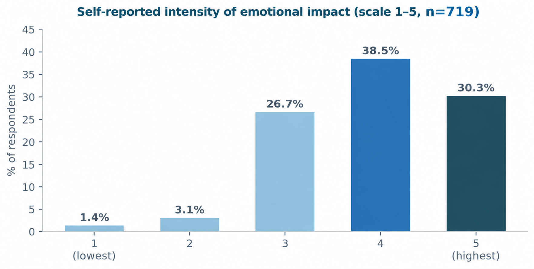
The most commonly reported emotional response to learning of an AL was concern for the patient (81.4%), followed by anxiety or distress (49.5%) and frustration or a sense of failure (48.5%). In contrast, fear of legal consequences (22.1%) and guilt or self–blame (21.6%) were less common, although they were associated with the highest levels of emotional distress (Fig. 2).
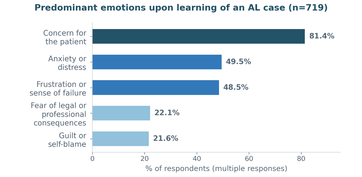
These findings suggest that, although surgeons’ primary concern when faced with AL is the patient’s clinical course, emotions related to self–blame and professional responsibility are associated with greater emotional distress.
Emotional impact varied according to respondent characteristics. Women more frequently reported high emotional impact than men (75.1% vs 66.7%; p=0.04), as well as higher rates of frustration or a sense of failure (64.3% vs 43.3%; p=0.001) and guilt or self–blame (30.3% vs 18.5%; p=0.001) (Fig. 3). Similarly, less experienced surgeons more frequently reported feelings of failure (61.8% vs 39.9%; p=0.001) and guilt (25.7% vs 18.8%; p=0.035). Colorectal surgeons also reported high emotional impact more frequently than other respondents (73.6% vs 66.2%; p=0.048), with no significant differences in the specific emotions reported. No significant differences in emotional impact were observed by country of residence (Argentina vs other countries; p=0.848).
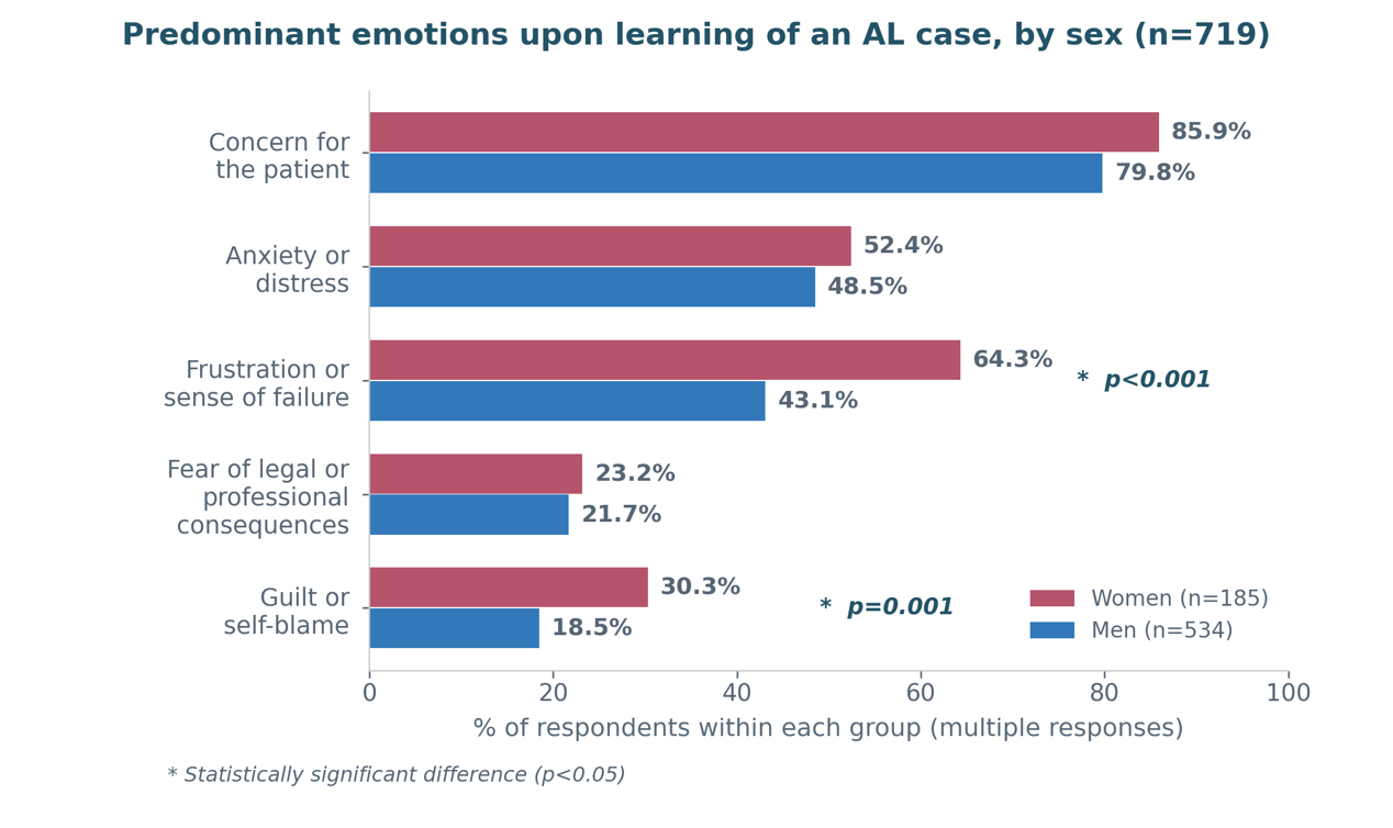
The impact of AL on respondents’ personal well–being outside the workplace was also assessed (Fig. 4). Persistent concern about the patient was the most frequently reported effect (82.5%), followed by difficulty disconnecting from work (41.0%), emotional exhaustion (38.9%), difficulty sleeping (25.7%), and irritability (19.6%). Only 5.6% of respondents reported no relevant changes.
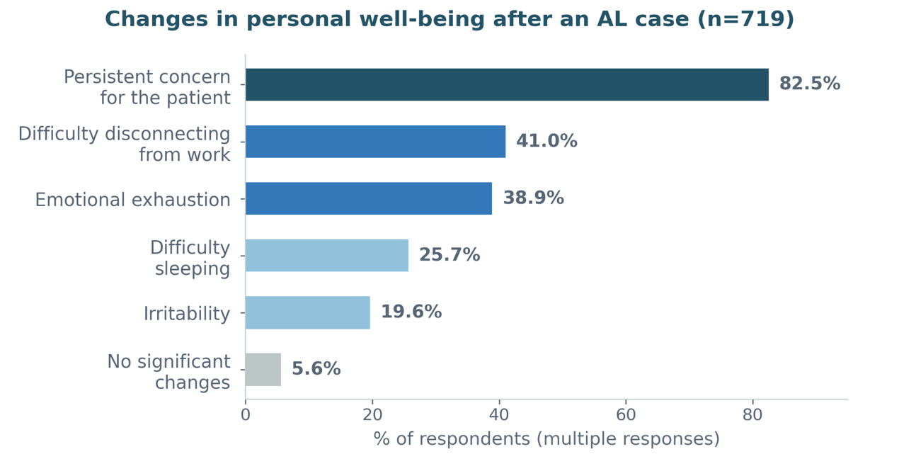
These changes in personal well–being also varied by respondent characteristics, although the patterns were not uniform. Women more frequently reported difficulty disconnecting from work (51.9% vs 37.3%; p=0.001) and emotional exhaustion (46.5% vs 36.3%; p=0.019) than men (Fig. 5), with no significant differences in persistent concern, difficulty sleeping, or irritability.
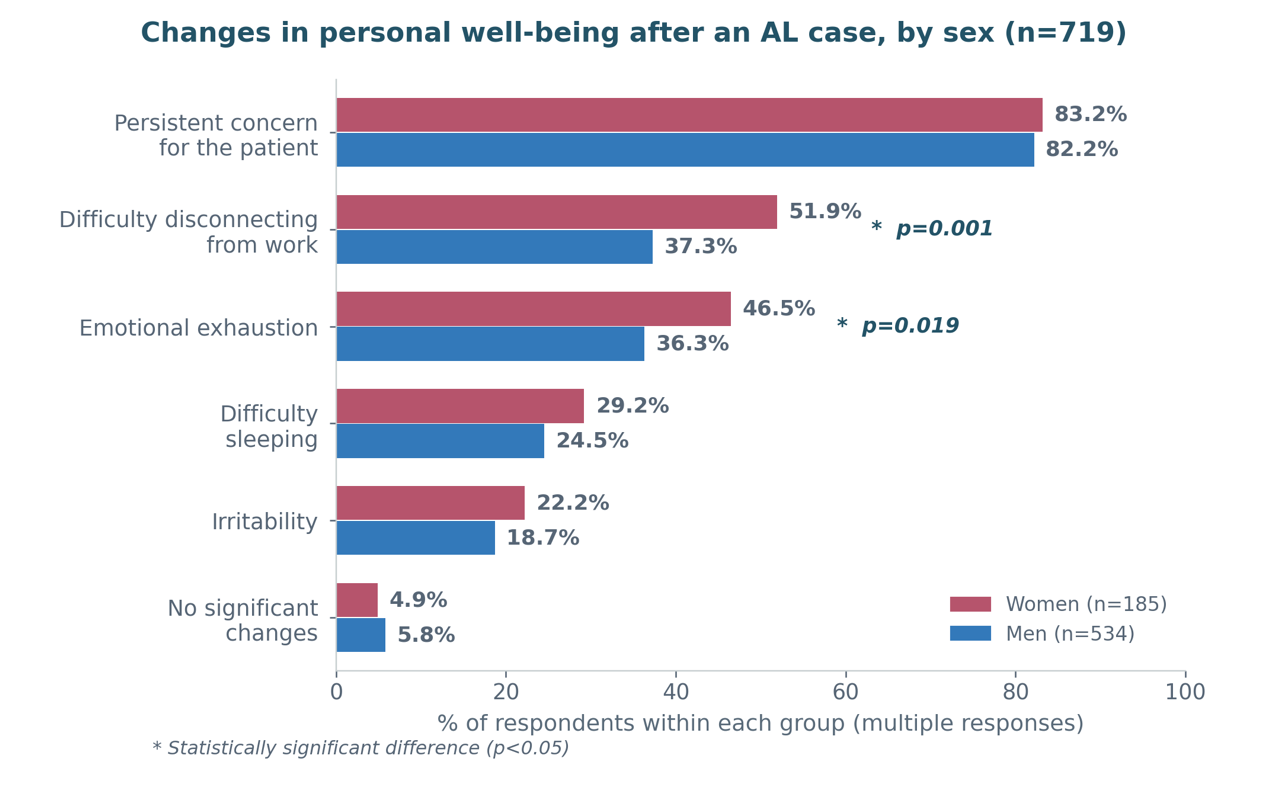
Less experienced surgeons also more frequently reported difficulty disconnecting from work (48.6% vs 36.0%; p=0.001). Notably, 2 outcomes showed the opposite pattern according to experience: persistent concern about the patient (85.6% vs 77.8%; p=0.009) and difficulty sleeping (29.2% vs 20.5%; p=0.011) were more frequently reported by more experienced surgeons. These findings suggest that although the overall emotional impact tends to be greater earlier in a surgical career, certain persistent effects may accumulate or manifest differently with increasing years in practice.
Finally, respondents from Argentina more frequently reported difficulty disconnecting from work than those from other countries (43.9% vs 29.4%; p=0.0012), with no significant differences in the other reported changes.
Changes in Clinical Practice
Nearly 2 in 3 surgeons reported modifying some aspect of their clinical or surgical practice after experiencing a major AL, highlighting the profound impact of this complication on professional behavior. Less experienced surgeons were significantly more likely than their more experienced counterparts to report changes in practice (68.4% vs 60.3%; p=0.034), suggesting that AL has a greater influence on professional practice earlier in a surgeon’s career. No significant differences were observed by sex or specialty.
The most common change after a major AL was not technical but communicational: nearly half of respondents reported devoting more attention to communication with patients and their families (45.5%). This was followed by greater caution when deciding to perform an anastomosis (38.8%) and a lower threshold for reoperation or drainage (34.5%) (Fig. 6). Women and less experienced surgeons were more likely to report placing greater emphasis on communication and seeking second opinions, whereas colorectal surgeons more frequently reported a more intervention–oriented approach, characterized by a lower threshold for reoperation.
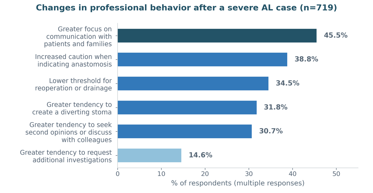
Overall, 43.9% of respondents reported taking steps to avoid similar situations after an episode of AL, most commonly by applying stricter selection criteria for high–risk patients (25%).
This approach was significantly less common among colorectal surgeons than among other surgeons (37.0% vs 46.3%; p=0.02), with no significant differences by sex or years of professional experience.
Relationship Between Emotional Impact and Changes in Practice According to Surgical Experience
The preceding findings may initially appear paradoxical: less experienced surgeons were more likely to modify their practice (68.4% vs 60.3%) and reported greater frustration and guilt, whereas more experienced surgeons were less likely to change their practice despite reporting more persistent distress in certain domains, including persistent concern about the patient (85.6% vs 77.8%; p=0.009) and difficulty sleeping (29.2% vs 20.5%; p=0.011) (see previous section, Fig. 5). However, an important distinction should be made: overall emotional impact, measured on a 5–point scale, did not differ significantly between the groups (69.1% vs 68.4% reporting high emotional impact; p=0.899). Rather, the predominant manifestations of distress appeared to vary across career stages.
To further explore this relationship, we examined whether the intensity of emotional impact was associated with the likelihood of changing clinical practice within each experience group. Among less experienced surgeons, the proportion who modified their practice was nearly identical among those reporting high and low emotional impact (68.5% vs 68.1%; p=1.00). This finding suggests that, early in a surgical career, changes in practice after a major AL may occur largely irrespective of the intensity of the emotional response. Among more experienced surgeons, there was a nonsignificant trend toward more frequent changes in practice among those reporting high emotional impact than among those reporting low emotional impact (63.1% vs 54.1%; p=0.099). These findings suggest that changes in practice may be a more consistent response to major AL among less experienced surgeons, whereas among more experienced surgeons, such changes may be more closely associated with the emotional impact of the event, potentially reflecting more established practice patterns that are less readily modified.
Professional Confidence and Medicolegal Concerns
Although AL has a substantial emotional impact, most surgeons did not report a marked loss of professional confidence. Only 10.7% reported a substantial effect on their self–confidence (ratings of 4 or 5 on a 5–point scale), whereas 59.1% rated the impact as 1 or 2 (Fig. 7).
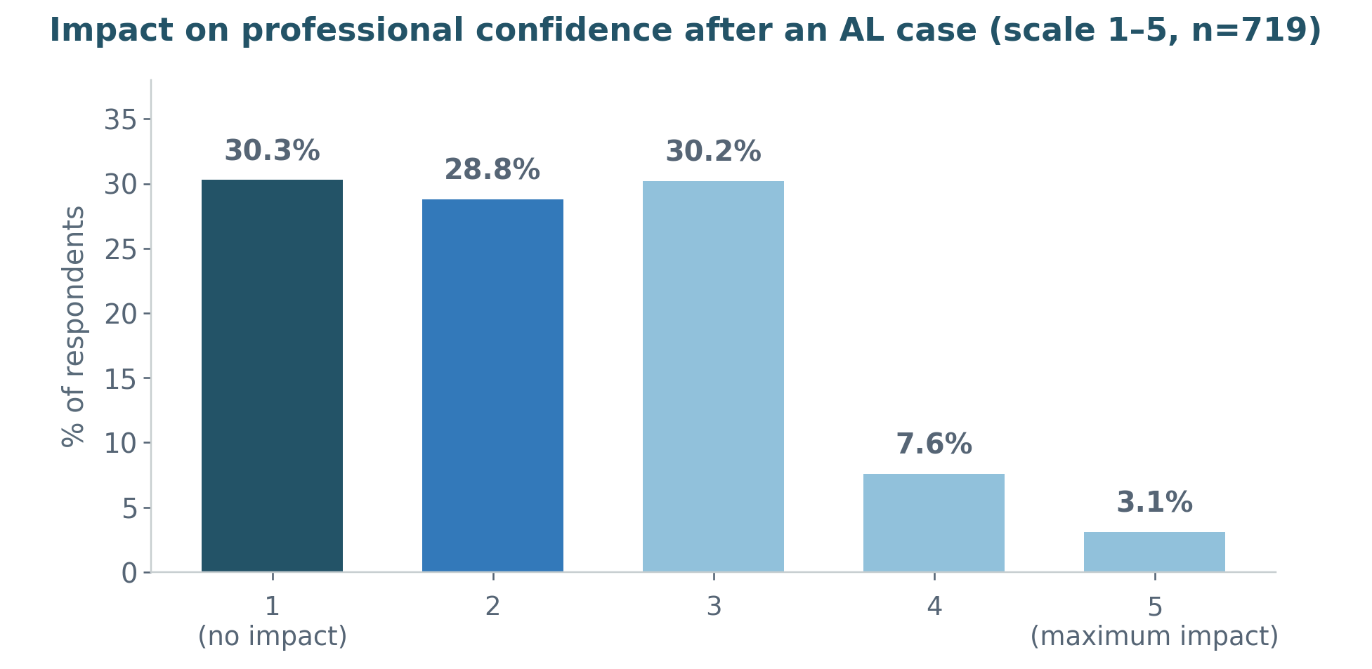
The impact on professional confidence was assessed across the full 5–point scale according to sex and surgical experience, rather than solely by the proportion of respondents reporting a high impact. Women reported significantly greater effects on professional confidence than men, with mean scores of 2.56 and 2.13, respectively (p<0.001) (Fig. 8).
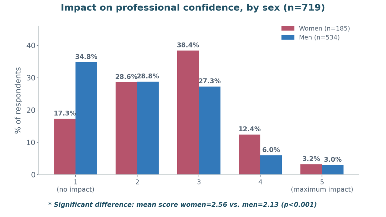
Conversely, a score of 1, indicating no impact on professional confidence, was reported by 34.8% of men compared with 17.3% of women. A significant difference was also observed according to surgical experience: surgeons with fewer than 11 years in practice had a mean score of 2.46, compared with 2.10 among more experienced surgeons (p<0.001) (Fig. 9). Notably, when the analysis was restricted to respondents reporting high impact (scores of 4–5), the difference between groups was not statistically significant (9.5% vs 12.5%; p = 0.252). However, the difference became significant when the full score distribution was considered, largely because of differences at the lowest end of the scale: 37.8% of more experienced surgeons reported no impact, compared with 19.1% of less experienced surgeons. Thus, the difference was driven primarily by the proportion reporting no impact rather than by the proportion reporting the highest levels of impact. No significant differences were observed according to colorectal specialization or country of residence.
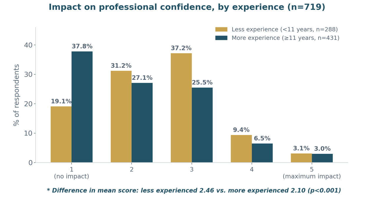
Regarding concerns about potential medicolegal consequences, 32.4% of respondents rated their level of concern as 4 or 5 on a 5–point scale, whereas 14.2% reported no concern (score of 1) (Fig. 10). Medicolegal concerns were similar across all groups analyzed, including sex, surgical experience, specialty, and country of residence.
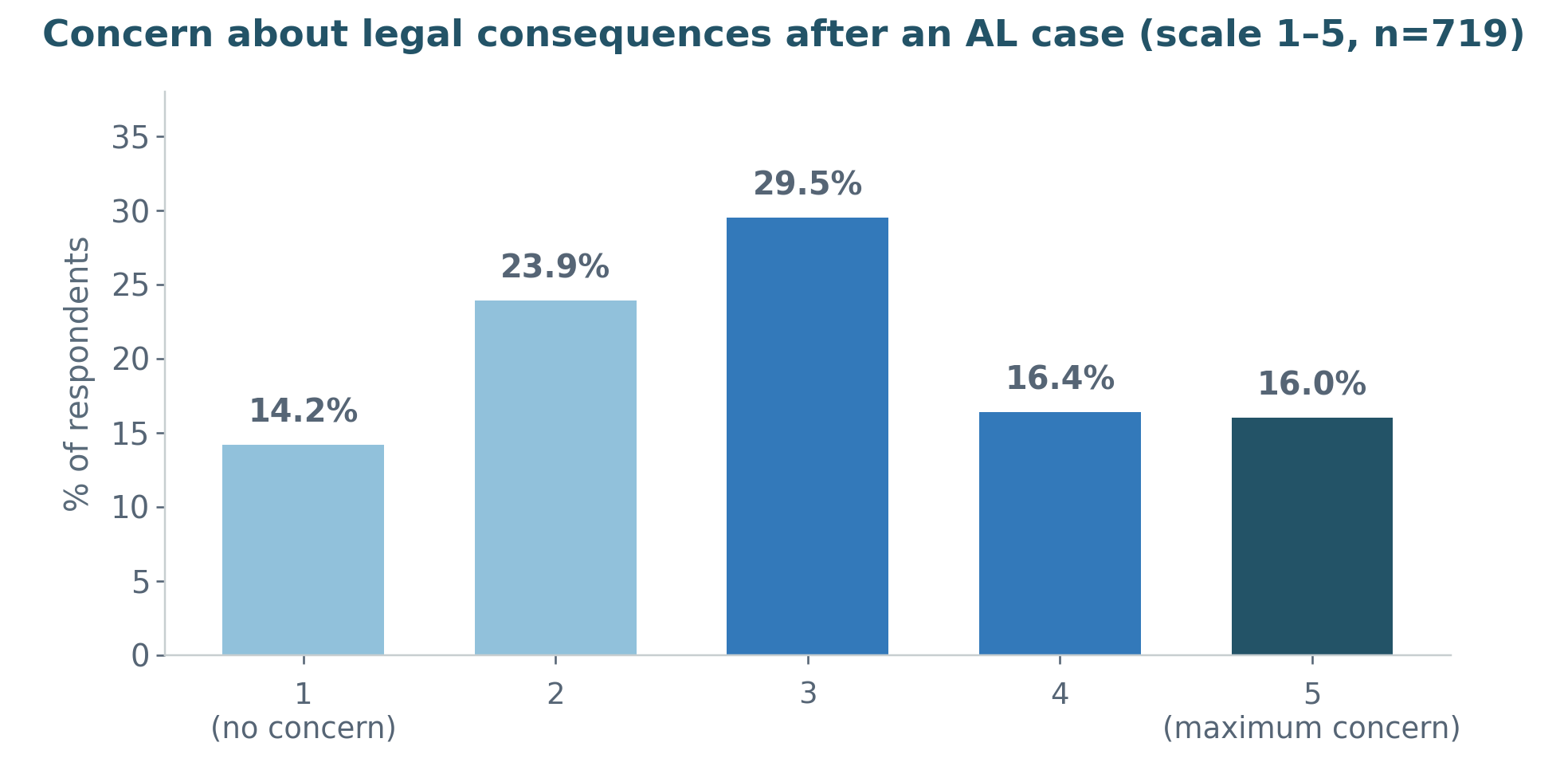
Colleagues were the most common source of emotional support after AL, with 68.3% of respondents reporting that they had discussed the event with a colleague.
Women and younger surgeons more frequently sought support from senior colleagues, mentors, and mental health professionals. Only 5.7% of respondents (n=41) reported that they had not felt there was an appropriate setting in which to discuss the case.
An important question is whether this subgroup experienced less emotional distress and therefore did not seek or need such a setting, or whether these surgeons were equally or more affected but lacked an adequate channel for support. The findings support the latter interpretation (Fig. 11). This subgroup did not report lower emotional impact than the remaining respondents (identical median scores on the 5–point scale; no statistically significant difference). However, these surgeons reported significantly greater impairment of professional confidence (mean score, 2.63 vs 2.22; p=0.018) and greater concern about potential medicolegal consequences (mean score, 3.39 vs 2.94; p=0.03) than those who had access to a setting in which to discuss the event.
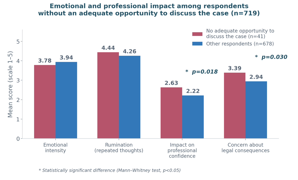
These findings suggest that the absence of a perceived appropriate setting for discussion is not associated with less emotional distress but may identify a particularly vulnerable subgroup characterized by greater erosion of professional confidence and heightened medicolegal concerns, without access to the support available to other colleagues. Given the small size of this subgroup (n=41), these findings should be interpreted cautiously and considered hypothesis–generating, warranting further investigation.
Finally, 5.4% of respondents reported having faced a medicolegal claim related to AL. Among these respondents, perceived communication problems with the patient or family were the most frequently reported factor associated with the claim, followed by delays in therapeutic intervention (reoperation, drainage, or diversion) and inadequate preoperative information or informed consent. Technical errors, in contrast, were less frequently identified as the primary cause.
No significant differences were observed according to sex, years of surgical experience, or level of specialization, likely owing to the small number of cases. These findings are consistent with those described above: from the surgeons’ perspective, communication–related factors play a more prominent role than the technical occurrence of AL itself, both in the circumstances leading to medicolegal claims and in subsequent changes in clinical practice after AL.
CONCEPTUAL SUMMARY
The survey findings indicate that AL is experienced by surgeons not merely as a technical complication but as an event with substantial professional, emotional, and behavioral consequences. When AL is suspected, clinical decision–making relies primarily on the patient’s clinical status and CT findings, although the use of rectal contrast remains limited despite its diagnostic value.
From an emotional standpoint, AL has a substantial impact on most respondents, characterized primarily by concern for the patient, anxiety, frustration, and, to a lesser extent, guilt or fear of medicolegal consequences. This impact varies across respondents, with women, less experienced surgeons, and colorectal specialists reporting greater effects in different domains.
Many surgeons modify their practice after a major AL, not only in technical aspects but particularly in communication. They devote more time to discussions with patients and families, exercise greater caution when considering anastomosis, and adopt a lower threshold for intervention, drainage, or seeking a second opinion. Among less experienced surgeons, these changes appear to occur almost automatically after the event, whereas among more experienced surgeons, they may be more closely related to the emotional impact of the individual case.
The survey also highlights the central role of communication. Communication problems with patients and families were the factor most frequently associated with medicolegal claims and, at the same time, the aspect of practice most commonly reinforced after AL. Finally, the absence of an appropriate setting in which to discuss the event identified a particularly vulnerable subgroup with greater impairment of professional confidence and greater concern about medicolegal consequences.
Taken together, these findings suggest that AL should be addressed not only as a surgical complication but also as a complex professional experience requiring emotional support, effective communication, and institutional mechanisms for supporting surgeons.
X. Conclusions
AL remains one of the most consequential, feared, and challenging complications of colorectal surgery. Despite advances in surgical techniques, the adoption of new technologies, the standardization of perioperative care, and a better understanding of risk factors, its incidence has remained relatively stable, underscoring its complex, multifactorial nature and the limitations of current preventive strategies. Anastomotic integrity depends on the interplay of patient–related factors, underlying disease, surgical technique, tissue perfusion, the inflammatory response, wound healing, and, increasingly, the intestinal microbiome.
Preventing AL requires a comprehensive assessment of each patient’s risk factors. Although no predictive scoring system can replace clinical judgment, risk stratification is essential when determining whether to perform a primary anastomosis, construct a diverting stoma, or defer restoration of intestinal continuity.
Intraoperatively, the fundamental principles remain unchanged: adequate perfusion of the bowel ends, a tension–free anastomosis, proper tissue apposition, and meticulous surgical technique. In high–risk patients, a diverting stoma should be considered a strategy to mitigate the clinical consequences of a potential AL rather than a measure that prevents the complication itself.
Diagnosing AL requires a high index of suspicion. Clinical assessment remains central, and any deviation from the expected postoperative course should alert the surgeon. Serum biomarkers, particularly CRP and procalcitonin, can complement clinical assessment, especially because of their NPV. However, no single biomarker can reliably confirm or exclude AL.
CT is the imaging modality of choice when AL is suspected, although it is not infallible. A normal CT scan does not exclude AL when clinical suspicion remains high. Intravenous contrast is essential, and rectal contrast, although underused in clinical practice, may improve diagnostic sensitivity in colorectal anastomoses by demonstrating contrast extravasation, one of the most specific radiologic findings of AL. Accordingly, imaging and laboratory studies should guide clinical decision–making but should never replace clinical assessment of the patient.
Management should be individualized according to hemodynamic status, anastomotic location, defect size, the presence of collections, viability of the bowel ends, and the extent of intra–abdominal contamination. In selected, hemodynamically stable patients, nonoperative management, PD, or endoscopic interventions may achieve source control while preserving the anastomosis. Conversely, sepsis, generalized peritonitis, ischemia of the bowel ends, extensive anastomotic defects, or hemodynamic instability warrant prompt surgical intervention aimed at source control and reducing morbidity and mortality.
Early reoperation is a critical determinant of outcome. In selected patients, relaparoscopy is a safe alternative that may be associated with lower morbidity and faster recovery. When the anastomosis remains viable and the defect is limited, anastomotic salvage with washout, drainage, and proximal diversion constitutes a reasonable strategy. In contrast, extensive defects, ischemia, or hemodynamic instability often necessitate anastomotic takedown and creation of an end stoma. Damage–control surgery should be reserved for critically ill patients, with priority given to rapid control of sepsis and early abdominal closure whenever feasible.
The consequences of AL extend well beyond the immediate postoperative period. This complication increases morbidity and mortality, prolongs hospitalization, raises healthcare costs, increases the likelihood of reoperation and permanent stoma formation, and impairs quality of life. In patients with cancer, AL may also delay or preclude adjuvant therapy and may be associated with worse oncologic outcomes. Furthermore, it is a major source of medicolegal claims, particularly when diagnostic or therapeutic delays or failures in communication occur.
Finally, the survey presented in this review provides a particularly valuable perspective: AL affects not only patients but also surgeons and the broader surgical team. The findings demonstrate a substantial emotional burden, with considerable proportions of respondents reporting concern for the patient, anxiety, frustration, guilt, and fear of medicolegal consequences. Many surgeons also reported modifying their practice after a major AL, particularly with respect to communication, decisions to construct an anastomosis, thresholds for reoperation or drainage, and seeking second opinions.
Taken together, this review highlights AL as a clinical, technical, biological, human, and institutional challenge.
Optimal management requires individualized prevention, early diagnosis, a stepwise treatment strategy, timely source control, effective communication with patients and families, and support for the surgical team. Rather than being viewed as an isolated complication, AL should be recognized as a critical event that challenges clinical decision–making, quality of care, surgical training, and the culture of support within healthcare teams.