Emiliano Caruso, Carla Peña Montecinos, Juan I. O'Brien, Cecilia Felipe Ramudo, Carlos A. Petrelli
General Surgery Department, Colorectal Surgery Section, Sanatorio Ipensa, La Plata, Buenos Aires, Argentina
Correspondence
Emiliano Caruso caruso95611@gmail.com
Received: April 4, 2026
Accepted: June 19, 2026
How to cite: Caruso E, Peña Montecinos C, O'Brien Juan I, Felipe Ramudo C, Petrelli CA. Endoluminal Negative Pressure Therapy as a Conservative Approach to Colorectal Anastomotic Leakage. Rev argent. De Coloproctologia. 2026;37(3). doi: 10.46768/v153m240
Keywords: anastomotic leakage; minimally invasive surgery; vacuum-assisted therapy; colorectal anastomosis
Anastomotic dehiscence following colonic resection is a potentially serious complication that affects patient morbidity and mortality, the number of reoperations, length of hospital stay, and the initiation of adjuvant cancer treatments.1,2
Vacuum-assisted closure (VAC) therapy is a locoregional management method consisting of controlled negative pressure to a wound bed to facilitate drainage and promote wound healing.3 This method has emerged as an alternative to conventional surgical treatments to preserve the anastomosis and facilitate healing of the surgical wound bed.1,4
VAC consists of a fenestrated polyurethane sponge attached to a drainage tube that connects to a continuous negative-pressure suction system (commercial VAC device or adjustable hospital-grade suction unit). Placement is guided by endoscopic equipment (colonoscope or rigid/flexible rectoscope).
VAC is indicated in clinically stable patients without diffuse peritonitis who have a localized anastomotic leak and a perianastomotic cavity accessible via the transanal route, ideally within 6 weeks of the index operation. It may also be applied following the formation of chronic perianastomotic cavities, such as a presacral sinus, which represents one of the most appropriate indications.
VAC is contraindicated in patients with generalized peritonitis, systemic sepsis, or hemodynamic instability; uncorrected coagulopathy or a high risk of bleeding; cavities that are inaccessible via the transanal route or associated with complex fistulas involving adjacent organs; active necrotic tissue that cannot be adequately debrided; or residual tumor that may preclude adequate wound healing.
Preprocedural assessment: Clinical evaluation of the patient, followed by contrast-enhanced computed tomography (CT) with rectal contrast or endoscopy to determine the extent, location, and accessibility of the cavity.
Preparation: Local enema or rectal cleansing; sedation/analgesia or anesthesia according to patient tolerance; and standard perineal preparation.
Initial irrigation and debridement: Under endoscopic guidance, the cavity is irrigated with saline, and fibrinous material or debris is removed. Hemostasis is achieved as needed.
Sponge placement: The polyurethane sponge is trimmed to fit the dimensions of the cavity and carefully introduced transanally into the perianastomotic cavity under direct endoscopic visualization. The sponge is secured to the drainage catheter, which is brought out through the anal canal.
Negative-pressure therapy: The catheter is connected to a negative-pressure system, typically at −75 to −125 mmHg, using continuous suction, with adjustments based on patient tolerance and the presence of bleeding.
Monitoring: Adequate sealing of the cavity is confirmed after connection to the suction system. Pain, bleeding, and drainage are monitored, and the volume and characteristics of the effluent are documented. Analgesia is provided as needed.
Sponge exchange: The sponge is removed and exchanged every 48–120 hours (typically every 3–5 days), with repeat irrigation, endoscopic assessment, and adjustment of sponge size according to the evolution of the cavity. Treatment is continued until the cavity progressively decreases in size and becomes filled with healthy granulation tissue.
Criteria for completion: Therapy is discontinued when endoscopy or contrast-enhanced CT with rectal contrast demonstrates complete resolution of the cavity, with a marked reduction in drainage and macroscopic or apparent closure of the defect. Once closure is confirmed, the system is removed and the patient continues with outpatient follow-up.
A 56-year-old man with rectal adenocarcinoma located 6 cm from the anal verge was initially staged as cT4aN1M0. He underwent neoadjuvant chemoradiotherapy followed by low anterior resection with a colorectal anastomosis located 4 cm from the anal verge and a diverting loop ileostomy. Final staging was yT3N0M0, reflecting an incomplete response to neoadjuvant treatment.
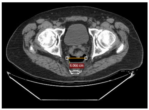
On postoperative day 12, the patient was readmitted with dehydration, nausea, fever, and leukocytosis. Abdominopelvic CT demonstrated a 6-cm perianastomotic collection containing an air-fluid level (Fig. 1). Rectosigmoidoscopy revealed a partial anastomotic defect involving more than 50% of the anastomotic circumference. Endoscopic irrigation of the surgical bed with saline solution was performed, and antibiotic therapy was initiated. The patient subsequently improved clinically, and follow-up CT demonstrated complete resolution of the perianastomotic collection.
Forty-five days after surgery, surveillance colonoscopy demonstrated persistent partial anastomotic dehiscence with a residual perianastomotic cavity measuring approximately 40 mm in diameter. The cavity had friable walls with adherent fibrin. In the absence of ongoing clinical signs of infection and given the persistent contained cavity, VAC was selected to promote cavity healing and preserve the colorectal anastomosis.
After rectal preparation with a saline enema (Enemol®), the cavity was irrigated under endoscopic guidance. A polyurethane sponge connected to a drainage catheter was then trimmed to fit the cavity and positioned endoscopically within the perianastomotic cavity. The catheter was connected to a continuous negative-pressure system set at −150 mm Hg (Fig. 2).
The patient remained hospitalized. The sponge was exchanged every 5 days, with cavity irrigation and endoscopic reassessment at each session. Three sponge exchanges were performed, with progressive reduction of the cavity and development of healthy granulation tissue. On treatment day 17, CT with rectal contrast demonstrated no contrast extravasation and no residual perianastomotic cavity. The patient reported no pain and experienced no complications related to VAC throughout treatment.
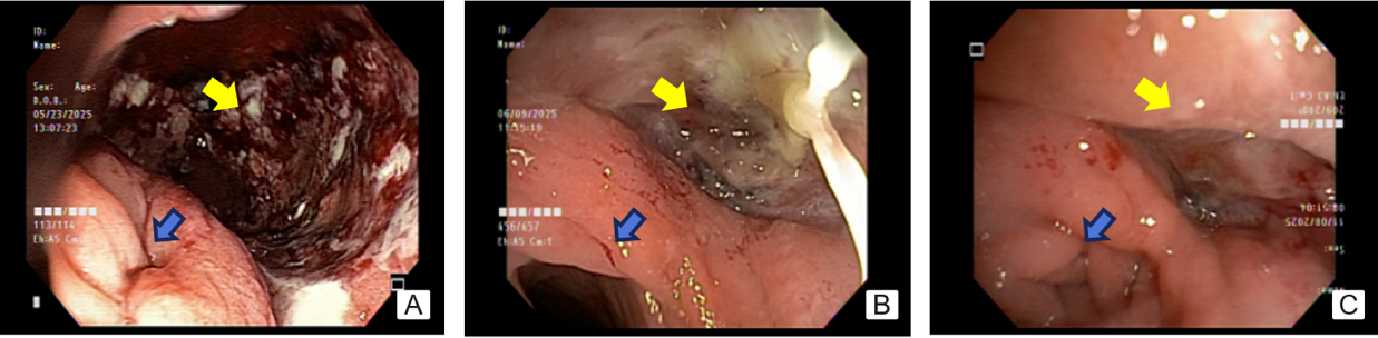
Thirty days after initiation of VAC, endoscopic examination confirmed complete healing of the anastomosis (Fig. 3). Given the satisfactory healing of the anastomosis, the diverting ileostomy was subsequently closed.
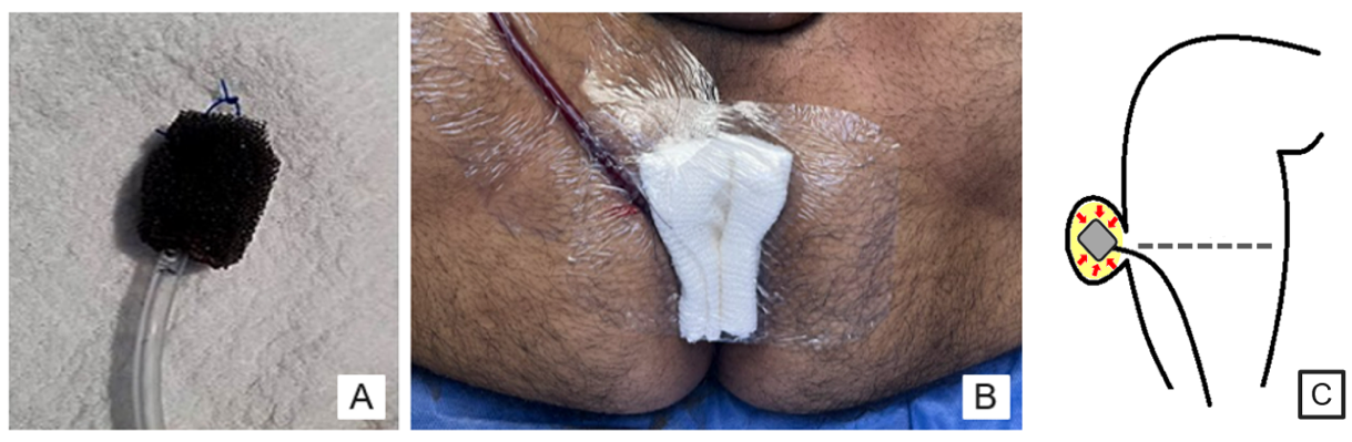
B. VAC system in place. C. Schematic illustration of VAC therapy within the cavity.
The reported incidence of anastomotic leakage following colorectal cancer surgery ranges from 3% to 19%, with associated mortality rates ranging from 1.7% to 16.4% across published series.1,2 One of the most common and characteristic manifestations of anastomotic leakage after low anterior resection is the development of a presacral sinus. This occurs when the anastomotic leak drains into the retrorectal space, resulting in a chronic cavity between the anastomosis and the presacral fascia that tends to epithelialize and persist without spontaneous closure. Several factors, including nutritional status, tumor height relative to the anal verge, prior radiotherapy, and operative time, may influence anastomotic healing.
Treatment selection depends on the patient’s overall condition, the presence and extent of any associated collection, and the location and integrity of the anastomosis. In patients with diffuse peritonitis and poor clinical status, conventional surgical management is generally preferred, including resection or takedown of the anastomosis with creation of a diverting stoma.⁵
Conversely, conservative approaches aimed at preserving the anastomosis are indicated in clinically stable patients with small defects. Although minimally invasive approaches can be effective in selected patients, healing may be prolonged and associated with an increased risk of anastomotic stricture.
Several treatment modalities have been described, including transanastomotic catheter drainage, percutaneous drainage, and endoscopic interventions such as clip placement and stent deployment across the defect.1,4
Negative-pressure therapy for the treatment of colorectal anastomotic leakage was first described in 2007, with favorable outcomes reported in the management of low rectal leaks.³ In recent years, this approach has gained increasing acceptance as a minimally invasive alternative to conventional treatment, particularly in selected patients with extraperitoneal leaks and well-defined presacral cavities.
VAC offers several potential benefits, including reduced drainage and edema, decreased bacterial burden, improved tissue perfusion, and enhanced granulation tissue formation. Negative-pressure therapy also promotes progressive reduction and eventual obliteration of the cavity to achieve defect closure and preserve the anastomosis, thereby potentially avoiding major surgical reintervention in selected patients. The negative pressure applied has ranged from 75 to 125 mmHg across published protocols, with serial sponge exchanges and cavity irrigation performed according to the clinical and endoscopic response.⁶ However, there is currently no universally accepted protocol regarding the optimal pressure, interval between sponge exchanges, or overall treatment duration. These parameters are generally individualized according to cavity characteristics, drainage volume, granulation tissue appearance, and findings on serial endoscopic assessments.
However, no universally accepted protocol currently exists regarding the optimal negative pressure, interval between sponge exchanges, or overall treatment duration. These parameters are typically individualized based on cavity characteristics, drainage volume, granulation tissue appearance, and findings on serial endoscopic assessments.
One aspect that remains controversial is the definition of treatment success. Currently, there is no consensus regarding the criteria used to determine resolution of anastomotic leakage. Some studies, such as CLEAN⁷ and the French GRECCAR group study,⁸ have used the absence of contrast extravasation on follow-up imaging or the endoscopic appearance of the anastomosis as criteria for treatment response. Other authors have used parameters such as reduction in cavity size, cavity closure with more than 90% granulation tissue, decreased wound drainage, or the absence of the need for additional surgical procedures.
Several observational studies and case series⁹ have reported widely variable rates of cavity closure. These outcomes appear to be significantly influenced by early initiation of therapy, particularly within 6 weeks after the index operation. There is also no consensus regarding the optimal duration of treatment, as treatment durations reported in the literature vary considerably.3,7
Several factors may influence the efficacy of negative-pressure therapy. Residual tumor, previous radiotherapy, nutritional status, and the presence or absence of a diverting stoma may all affect treatment outcomes.
Despite its advantages, this strategy has several limitations. The need for multiple endoscopic sponge exchanges, the cost associated with commercially available devices, and treatment-related bleeding, discomfort, or pain of varying intensity may limit treatment tolerance and continuity.
Chronic complications have also been described, including fistula formation (enteric, colovesical, colovaginal, ileal, or urethral fistulas), as well as anastomotic stricture.³
Vacuum-assisted closure therapy is an effective minimally invasive option for the conservative management of colorectal anastomotic leakage in selected patients, particularly those with localized leaks and adequate infection control. It may preserve the anastomosis while avoiding major surgical reintervention.
Vacuum-assisted closure therapy is an effective minimally invasive option for conservative management of colorectal anastomotic leakage in selected patients, particularly those with localized leaks and adequate infection control, with the potential to preserve the anastomosis and avoid major surgery. Additional benefits include reducing the need for a permanent stoma, improving quality of life, and enabling earlier resumption of adjuvant therapy. Although published results are promising, the available evidence remains limited by study heterogeneity and the lack of standardized treatment protocols and definitions of therapeutic success. The decision to use VAC therapy should be individualized according to the characteristics of the leak, the patient’s overall condition, and oncologic and functional considerations.
Author Contributions:
EC: Conceptualization, Methodology, Writing – original draft, Supervision
CPM: Data curation, Research, Writing – review and editing
JIOB: Research, Resources, Data Collection
CFR: Validation, Writing – review and editing
CAP: Project administration, Supervision, Validation
Conflict of interest statement: None.
Funding: None.
Data availability statement: The data are publicly available.
ORCIDs:
Emiliano Caruso: https://orcid.org/0000-0001-7286-763X
Carla Peña Montecinos: https://orcid.org/0009-0004-8731-827X
Juan Ignacio O’Brien: https://orcid.org/0009-0007-3341-5930
Cecilia Felipe Ramudo: https://orcid.org/0009-0004-2790-1535
Carlos Alberto Petrelli: https://orcid.org/0009-0002-0664-7836
Blumetti J, Chaudhry V, Cintron JR, Park JJ, Marecik S, Harrison JL, et al. Management of anastomotic leak: lessons learned from a large colon and rectal surgery training program. World J Surg. 2014;38:985-91. doi: 10.1007/s00268-013-2340-y.
Bakker IS, Grossmann I, Henneman D, Havenga K, Wiggers T. Risk factors for anastomotic leakage and leak-related mortality after colonic cancer surgery in a nationwide audit. Br J Surg. 2014;101:424-32; discussion 432. doi: 10.1002/bjs.9395.
Weidenhagen R, Gruetzner KU, Wiecken T, Spelsberg F, Jauch KW. Endoscopic vacuum-assisted closure of anastomotic leakage following anterior resection of the rectum: a new method. Surg Endosc 2008; 22: 1818-25. doi: 10.1007/s00464-007-9706-x.
Phitayakorn R, Delaney CP, Reynolds HL, Champagne BJ, Heriot AG, Neary P, et al. Standardized algorithms for management of anastomotic leaks and related abdominal and pelvic abscesses after colorectal surgery. World J Surg 2008; 32: 1147-56. doi: 10.1007/s00268-008-9468-1.
Lim M, Akhtar S, Sasapu K, Harris K, Burke D, Sagar P, et al. Clinical and subclinical leaks after low colorectal anastomosis: a clinical and radiologic study. Dis Colon Rectum. 2006;49(10):1611-9. doi: 10.1007/s10350-006-0663-6.
Popivanov GI, Mutafchiyski VM, Cirocchi R, Chipeva SD, Vasilev VV, Kjossev KT, et al. Endoluminal negative pressure therapy in colorectal anastomotic leaks. Colorectal Dis 2020; 22: 243-53. doi: 10.1111/codi.14754.
Borstlap WAA, Musters GD, Stassen LPS, van Westreenen HL, Hess D, van Dieren S, et al. Vacuum-assisted early transanal closure of leaking low colorectal anastomoses: the CLEAN study. Surg Endosc 2018; 32: 315-27. doi: 10.1007/s00464-017-5679-6.
Abdalla S, Cotte E, Epin A, Karoui M, Lefevre JH, Berger A, et al. Short-term and long-term outcome of endoluminal vacuum therapy for colorectal or coloanal anastomotic leakage: results of a nationwide multicenter cohort study from the French GRECCAR Group. Dis Colon Rectum 2020; 63: 371-80. doi: 10.1097/DCR.0000000000001560.
Mahendran B, Rossi B, Coleman M, Smolarek S. The use of Endo-SPONGE® in rectal anastomotic leaks: a systematic review. Tech Coloproctol 2020; 24: 685-94. doi: 10.1007/s10151-020-02200-1.