Marta Moreno Ballesteros, Macarena Tejero García, Daniel Sánchez Peláez
Mérida Hospital, Department of General and Gastrointestinal Surgery
Correspondence
Marta Moreno marta.morenob@salud-juntaex.es
Received: 17-03-2026
Accepted: 19-06-2026
How to cite: Moreno M, Sánchez Peláez D, Tejero García M. Perianal Fistula as a Late Manifestation of Acute Diverticulitis: A Clinical and Surgical Analysis. Rev. argent. coloproctología 2026;37(3). doi: 10.46768/qezadv67
ABSTRACT
A colocutaneous fistula is a rare complication of diverticular disease, and its extension into the perianal region is exceptionally uncommon. We report the case of a 64-year-old man with a 1-year history of a perianal fistula following a perianal abscess, without a history of inflammatory bowel disease. Endoanal ultrasonography demonstrated an intersphincteric fistulous tract, although the internal opening could not be identified. Pelvic magnetic resonance imagin g (MRI) revealed a fistulous tract originating near the sigmoid colon and extending pararectally toward the levator ani muscle and left ischioanal fossa. Computed tomography demonstrated a complex fistula associated with perforation of a sigmoid diverticulum. The patient underwent laparoscopic sigmoidectomy with primary colorectal anastomosis. His postoperative course was uneventful, with no complications. This case highlights the value of pelvic MRI in delineating the anatomy and extent of complex fistulizing diverticular disease and in facilitating surgical planning. Laparoscopic sigmoidectomy with primary anastomosis may represent a feasible and safe approach in carefully selected patients.
Keywords: perianal fistula; colocutaneous fistula; acute diverticulitis; complicated diverticular colopathy; laparoscopic sigmoidectomy
A perianal fistula is an abnormal connection between the glandular epithelium of the anal canal and the perianal skin, most commonly representing a chronic sequela of a perianal abscess.1 Its true prevalence is difficult to determine, as many anal symptoms may be mistakenly attributed to hemorrhoidal disease Approximately 15% to 38% of patients with a perianal abscess subsequently develop a fistula.2
The most common etiology is cryptoglandular infection, resulting from obstruction and infection of the excretory ducts of the anal glands. The infectious process typically progresses along the path of least resistance through the intersphincteric space,2 with subsequent epithelialization of the fistulous tract leading to the formation of a mature fistula.
A second major etiologic group comprises fistulas associated with Crohn’s disease, in which the underlying pathogenic mechanism is transmural intestinal inflammation rather than cryptoglandular infection.3 Perianal fistulas may also occur secondary to obstetric trauma, radiation therapy, or infectious diseases such as tuberculosis and lymphogranuloma venereum, which may present with perianal masses.
Acute diverticulitis is a common condition in Western countries, with an incidence that increases with age and a higher prevalence among men.²
Among its most significant complications is fistula formation, which occurs in approximately 27% of surgically treated patients. The most common locations are colovesical, colovaginal, and coloenteric fistulas.3
A colocutaneous fistula is a rare complication of diverticular disease, occurring in fewer than 7% of cases. It may develop as a consequence of acute diverticulitis complicated by localized perforation and pericolic abscess formation, which subsequently establishes a fistulous tract to the skin or adjacent structures.1 In cases involving the pelvis—although uncommon—the inflammatory and infectious process may dissect through the pararectal planes and extend toward the perineum or perianal region.
A 64-year-old man was referred for evaluation of a 1-year history of a perianal fistula that had developed after a perianal abscess. He reported no abdominal pain, fever, or urinary symptoms during the course of the disease. He denied a history of previous abscesses or a personal or family history of inflammatory bowel disease.
Perianal examination in the lithotomy position revealed an external opening (EO) at the 3 o’clock position, 5 cm from the anal verge. After probing and instillation of saline solution, no communication with the anal canal was identified. Digital rectal examination did not reveal an internal opening (IO).
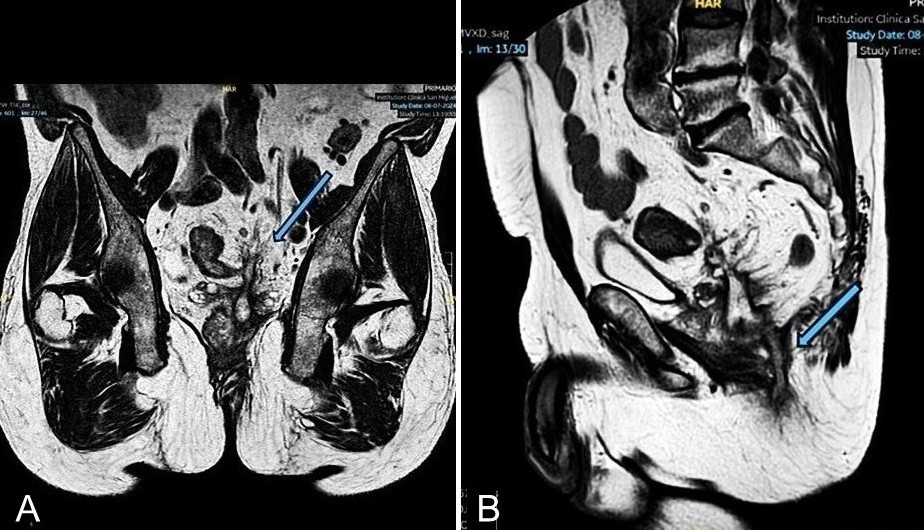
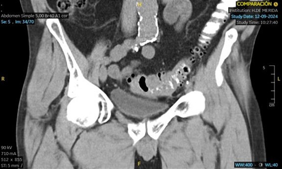
Endoanal ultrasonography revealed a curvilinear intersphincteric tract originating at the 3 o’clock position, with no identifiable IO. Pelvic magnetic resonance imaging (MRI) was therefore performed and demonstrated a fistulous tract adjacent to the sigmoid colon, extending in a pararectal course toward the levator ani muscle, with an associated abscess in close proximity (Fig. 1A). The distal portion of the tract extended to the left ischioanal fossa (Fig. 1B). Colonoscopy showed no evidence of active inflammatory disease. Abdominal and pelvic computed tomography (CT) demonstrated a complex fistula secondary to a perforated sigmoid diverticulum (Fig. 2).
A laparoscopic sigmoidectomy was planned, with preoperative left ureteral catheterization. Intraoperatively, a localized inflammatory process involving the sigmoid colon was identified, with dense adhesions to both small-bowel loops and the left paracolic region. These adhesions were carefully lysed. During dissection, a fistulous tract originating from the left anterolateral aspect of the sigmoid colon was identified. The tract extended into the extraperitoneal space and was densely adherent to the left seminal vesicle. Dissection of the fistulous tract was attempted; however, despite left ureteral catheterization, the inability to safely delineate the fistulous tract and its relationship to the ureter prompted conversion to open surgery to minimize the risk of ureteral injury and other intraoperative complications.
Following conversion to open surgery, the sigmoid colon was fully mobilized to facilitate a primary colorectal anastomosis. During dissection, dense inflammatory adhesions resulted in inadvertent splenic decapsulation, requiring splenectomy for hemostatic control. A midline supra- and infraumbilical laparotomy was subsequently performed to optimize exposure of the surgical field. The fistulous tract was identified and carefully dissected from the left seminal vesicle before being resected with electrocautery. Finally, the fistulous tract was curetted through the EO, completing a partial fistulectomy.
Histopathologic examination confirmed the presence of tissue consistent with a chronic fistulous tract.
In the immediate postoperative period, the patient developed a surgical-site seroma (Clavien-Dindo grade I). On postoperative day 20, he was readmitted with an intra-abdominal collection secondary to omental infarction, which was managed with percutaneous drainage. This resulted in a 19-day prolongation of the hospital stay.
Most colocutaneous fistulas arise as a complication of intestinal resection for diverticulitis⁴ or following percutaneous drainage of a diverticular abscess.⁴⁻⁸ Occasionally, sigmoid-perianal fistulas may develop spontaneously, through a specific anatomical pathway of spread.
Patients may report previous episodes of abdominal pain, nausea, or diarrhea, although some remain asymptomatic, as in the present case. In 2015, Kumar and Diaz⁵ reported the case of a female patient in whom a perianal abscess was the initial manifestation of diverticulitis.
Physical examination usually reveals a recurrent⁹ or persistent perianal abscess or fistula.5,10
The diagnosis of a perianal or cutaneous fistula secondary to diverticulitis poses a clinical challenge, given the extreme rarity of this complication and its atypical presentation, which frequently delay recognition.4,8,9 Contrast-enhanced CT of the abdomen and pelvis is essential for assessing the extent of disease and identifying acute complications.⁵ MRI is particularly useful because for delineating fistulous tracts and their anatomical relationship to adjacent structures.¹⁰
Diverticular fistulas rarely close spontaneously and usually require surgical intervention.9,10 Initial management of a perianal abscess arising from perforated acute diverticulitis consists of drainage. Definitive treatment requires bowel resection and excision of the fistulous tract, typically several weeks to months after control of the infection and resolution of the acute inflammatory process.
There is currently no established consensus regarding the optimal surgical approach, largely due to the high technical complexity of these procedures. Dense adhesions, fibrosis, and distortion of the anatomical planes may significantly hinder the identification and dissection of the involved structures, increasing the risk of visceral injury. In this context, laparoscopic surgery has emerged as a safe and feasible alternative for the treatment of complicated diverticular disease allowing sigmoidectomy and fistulotomy. Several studies also suggest potential advantages over the open approach, including less blood loss, shorter hospital stays, and, in certain scenarios, a reduction in surgical time.⁸
The open approach remains a valid option, particularly in patients with locally advanced disease or in centers with limited experience in laparoscopic surgery for complex fistulas⁴. Conversion to laparotomy should likewise be regarded as a safety strategy rather than a failure of the minimally invasive approach, particularly when extensive fibrosis, dense adhesions, or obliteration of the anatomical planes compromise safe dissection.4,8
The standard of care is to perform resection with primary anastomosis in a single stage,¹ as this approach has been shown to be safe and feasible in the absence of ongoing infection.² Although a laparoscopic Hartmann procedure with temporary end colostomy and subsequent restoration of intestinal continuity has been reported,¹⁰ this strategy appears to be exceptional and should be reserved for patients in whom local or systemic conditions preclude primary anastomosis.
Most published cases describe favorable clinical outcomes, with a low incidence of postoperative complications.7,8 Among the complications reported, pneumonia appears to be one of the most common, whereas anastomotic leaks have not been reported.5,6 Nevertheless, the available evidence is predominantly observational and is based on a limited number of published cases.
Although perianal fistulas secondary to acute diverticulitis are rare, early recognition and accurate characterization with magnetic resonance imaging can facilitate appropriate surgical planning. In selected patients, laparoscopic resection with primary anastomosis is a safe and feasible treatment option.
Author Contributions:
MMB: Conceptualization, Methodology, Investigation, Formal analysis, Data curation, Writing – Original Draft, Visualization.
MTG: Investigation, Resources, Supervision, Writing – Review & Editing.
DSP: Writing – Review & Editing, Supervision.
Conflict of interest statement: None.
Funding: None.
Data availability statement: The data are publicly available.
ORCIDs:
Marta Moreno Ballesteros: https://orcid.org/0009-0002-2339-2273
Daniel Sánchez Peláez: https://orcid.org/0000-0002-4116-6686
Macarena Tejero García: https://orcid.org/0009-0001-2307-7102
Abcarian H. Anorectal infection: abscess-fistula. Clin Colon Rectal Surg. 2011;24(1):14-21. doi: 10.1055/s-0031-1272819.
Cooper CR, Keller DS. Perianal fistulas. Dis Colon Rectum. 2020;63(2):129–32.
Parrilla Paricio P, Landa García JI, eds. Cirugía AEC: manual de la Asociación Española de Cirujanos. Madrid: Ed. Médica Panamericana; 2010.
Amor IB, Kassir R, Bachir E, Katharina H, Debs T, Gugenheim J. Perforated diverticulitis of the sigmoid colon revealed by a perianal fistula. Int J Surg Case Rep. 2015;8C:73–5.
Kumar K, Diaz P. Rare case of sigmoid-perianal fistula due to sigmoid diverticular disease: Report of a case and review of literature. J Med Cases. 2015;6(3):122–4.
Bakopoulos A, Tsilimigras D, Syriga M, Koliakos N, Ntomi V, Moris D, et al. Diverticulitis of the transverse colon manifesting as colocutaneous fistula. Ann R Coll Surg Engl. 2018;100(8):e191–3. doi:10.1308/rcsann.2018.0130.
Charalabopoulos A, Misiakos E, Macheras A. Colocutaneous fistula complicating sigmoid diverticulitis. Int J Surg Case Rep. 2011;2(5):68–70.
Hidaka E, Nakahara K, Maeda C, Takehara Y, Ishida F, Kudo S-E. Laparoscopic surgery for sigmoidocutaneous fistula due to diverticulitis: A case report: Surgery for sigmoidocutaneous fistula. Asian J Endosc Surg. 2015;8(3):340–2.
Ayoubi S, Chen M, Ravindran P, Gibson K. Ischio-anal abscess as a first presentation of complicated diverticular disease: Images for surgeons. ANZ J Surg. 2020;90(1–2):169–71.
Schmidt E, Corbitt M, Kulendran K, Ruggiero B. Fistulating diverticular disease masquerading as a peri-anal abscess: a laparoscopic approach to management. J Surg Case Rep. 2021;2021(11):rjab483. doi: 10.1093/jscr/rjab483