Giant solitary diverticulum of the sigmoid
colon
Giant solitary diverticulum of the sigmoid
colon

Fernando Fernández López1,
María Jesús Ladra González1, Jesús Paredes Cotoré1,
Manuel Bustamante Montalvo2
Complexo Hospitalario Universitario de Santiago, Hospital Clínico
Universitario de Santiago, Santiago de Compostela, España
1 Servicio de Cirugía General y Digestiva. Unidad de Coloproctología.
2 Jefe de Servicio de Cirugía General y Digestiva.
ABSTRACT
Solitary giant diverticulum of the sigmoid colon is a very
rare entity that can present as acute abdomen or with chronic nonspecific
abdominal discomfort. The most important complications are perforation,
obstruction and volvulation. Imaging studies show the characteristic “balloon
sign”, a large, smooth-walled gas-filled cyst. Treatment is surgical resection
of the affected colonic segment. We present the case of a patient with
recurrent episodes of diverticulitis that resolved with surgical resection.
Keywords: giant solitary diverticulum, sigmoid
colon, balloon sign, colonic resection
INTRODUCTION
Solitary
giant diverticulum of the sigmoid colon is a very rare entity that can present
as an acute abdomen or with chronic nonspecific abdominal discomfort
reminiscent of typical uncomplicated acute diverticulitis.1 The most
important complications of a giant diverticulum are perforation, obstruction or
volvulation. Both an abdominal x-ray and a barium enema show a large gas-filled
cystic image (balloon sign), with regular edges and a smooth wall,2
although computed tomography is the diagnostic test of choice. Treatment
consists of en-bloc resection of the compromised colon segment.3
Depending on the pathological appearance, these diverticula are classified as:
type 1 or pseudodiverticulum (pulsion type); type 2 or inflammatory type, and
type 3 or true diverticulum (contains all layers of the intestinal wall).4
CASE
A
47-year-old female patient with a history of multiple consultations for
episodes of chronic abdominal pain consulted due to exacerbation of cramping
pain.
The abdominal x-ray showed an image of intra-abdominal gas, similar to a
balloon. An abdominal CT scan reported a “large air bubble” in the left abdomen
with no identifiable origin (Fig. 1).
Once pneumoperitoneum was ruled out, the study was completed with a colonoscopy
that showed no findings of interest. The same intra-abdominal gas configuration
was observed in the barium enema (Fig. 2).
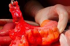
The patient underwent laparoscopic surgery, although
it had to be converted to open surgery due to adhesions and a severe
inflammatory process next to the sigmoid colon and angle of Treitz as a
consequence of recurrent acute episodes. A large solitary sigmoid diverticulum
was identified (Fig. 3).
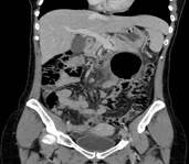
Figure 1. CT scan showing the "balloon sign," a large,
thin-walled gas bubble without continuity with the lumen of the colon.
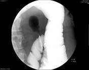
Figure 2. Barium enema.
The narrow lumen of the diverticular neck prevents passage of contrast into
the gas-filled diverticulum.
Despite having a narrow orifice, the base was very
wide, so it was decided to perform a sigmoidectomy with an end-to-end
colo-colonic anastomosis. The postoperative period was uneventful and the
patient was discharged on the sixth postoperative day.
The
pathological examination reported giant solitary colonic diverticulum (>5
cm), with signs of chronic inflammation and presence of foreign body giant
cells, without evidence of malignancy, type 2 according to the McNutt classification.4
DISCUSSION
A
giant diverticulum is defined as an air-filled cystic diverticulum of more than
4 cm in maximum diameter.1 Although its etiology is not clear, some
authors propose a valvular mechanism of colonic gas trapping that is
self-perpetuating with inflammatory episodes.5 Although the first
described case of solitary air cyst dates back to 1943, a small number of cases
have been published that describe this clinical entity with various names:
"giant gas cyst", "giant sigmoid diverticulum", "giant
colonic diverticulum" or "intestinal gas cyst".
A
giant diverticulum can present with a variety of signs and symptoms, ranging
from an incidental finding in an asymptomatic patient, to presentation as an
abdominal tumor or even an acute abdomen secondary to perforation.
The
main etiopathogenic mechanism described is the valvular effect of the muscular
layer that narrows the communication of the pseudodiverticulum of mucosa and
submucosa with the intestinal lumen due to the inflammatory process.
The diagnosis is suspected with a plain abdominal x-ray and confirmed with a
computed tomography scan. Both tests show the classic “balloon sign”. The
barium enema confirms the diagnosis when there is communication between the
diverticulum and the lumen of the colon, which only occurs in 25% of cases.
Sometimes, this study can precipitate perforation of the diverticulum.
Sigmoidoscopy rarely provides information.
The treatment of choice is resection of the affected colonic segment, with
anastomosis as long as there are no local complications such as perforation or
abscess. In these cases, a Hartmann’s procedure could be performed.
CONCLUSION
We
present the case of a giant solitary colon diverticulum type 2 (inflammatory).
It should be suspected in the presence of a balloon-shaped radiological image.
Its treatment is surgical to avoid complications such as the formation of
abscesses or free perforation.
REFERENCES
1. Nigri G, Petrucciani N, Giannini G, Aurello P,
Magistri P, Gasparrini M, et al. Giant colonic diverticulum: clinical
presentation, diagnosis and treatment:
systematic review of 166 cases. World J
Gastroenterol. 2015; 21:360-68.
2. Thomas S, Peel RL, Evans LE, Haarer KA. Best cases
from the AFIP: Giant colonic diverticulum. Radiographics. 2006; 26:1869-72.
3, Kam JC, Doraiswamy V, Spira RS. A rare case
presentation of a perforated giant sigmoid diverticulum. Case Rep Med.
2013;2013:957152.
4. McNutt R, Schmitt D, Schulte W. Giant colonic
diverticula-three distinct entities. Report of a case. Dis Colon Rectum. 1988;
31:624-28.
5. Salazar-Ibargüen J, Escárcega RO, Pérez
Chávez G. Giant sigmoid colon diverticulum. Dig Surg. 2007; 24:17-8.